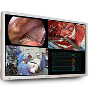
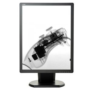
What Are Diagnostic Imaging Systems?
Diagnostic Imaging Systems are a vital component of clinical decision-making, and they are commonly used in many hospitals and health centers. Imaging provides physicians a tool not only to diagnose injuries or illness, but also to plan and monitor the course of needed treatments.
What Is Diagnostic Imaging?
Diagnostic medical imaging involves specific techniques that obtain images from inside the body. This technology provides detailed visualizations that include any abnormalities in structure or function. A diagnostic radiologist is a physician who is specially trained in the interpretation of these images to diagnose illness or injury.
What Is It Used For?
Diagnostic medical imaging allows physicians to visualize activities and structures inside the body. The images help diagnose the cause of symptoms or identify signs of a health condition. Diagnostic imaging also provides capabilities to confirm illnesses or monitor how well a patient is responding to medical treatment or intervention.
Types of Diagnostic Imaging
- An MRI scan can detect tumors, injuries, lesions, and infections. One benefit of this type of diagnostic imaging system is that it uses a powerful magnet (not radiation) to obtain a 3D image of organs and tissues inside the body of the patient. This scan can be used to examine the spinal cord, brain, joints, breast tissue, abdomen, or liver for injuries or abnormalities like cysts or tumors. An MRI exam can be ordered with contrast and generally takes about 30 to 60 minutes.
There are four types of MRI machines:
- True open: This is open on all sides; beneficial for individuals who are claustrophobic.
- Closed: A traditional tube is one in which a patient lies down inside for the images.
- Wide bore: This resembles a closed MRI, but with a wider middle opening.
- 3T MRI: This type is more advanced than traditional MRI and it takes less time. The high-resolution images enable the radiologist to determine the severity of a patient’s condition.
- MRA Scans (magnetic resonance angiogram) provide very detailed images of the blood vessels to identify issues that lead to reduced blood flow. Physicians use MRAs to look for calcium deposits, aneurysms, inflammation, or clots within the blood flow that may narrow or occlude vessels. In some cases, they may order a contrast dye to get a better definition of the scan’s images. MRA tests are typically used on the legs, neck, brain, or kidneys. In many cases, an MRA scan can detect more information than x-rays, ultrasounds, or CT scans.
- Computed tomography (CT) or computerized axial tomography (CAT) scans are frequently used to quickly examine individuals who may have internal injuries from trauma. CT scans are commonly used to evaluate the spine, brain, abdomen, neck, and chest. A CT scan can detect bone and muscle disorders, masses, tumors, injuries, and internal bleeding. This test combines a string of X-ray images taken at multiple angles to generate a cross-sectional slice of blood vessels and soft tissues. While CT scans use low doses of radiation, they’re still relatively non-invasive and safe.
A CT scan can be used to visualize nearly all body parts, including:
- Head: Check for stroke, masses, and other abnormalities
- Chest: Provide more detail into abnormalities as needed after an x-ray
- Neck: Look for enlarged glands or lymph nodes and lumps.
- Spine: Detect problems like spinal canal narrowing, a herniated disc, or fractures
- Sinus: Detect and diagnose obstructions or sinus disease
- Pelvis/ Abdomen: Check organs and diagnose unexplained pain
- Ultrasound- This technology produces real-time images onto a computer monitor as the technician moves the transducer over an area to show the structure and movement of internal organs or blood flow. It is often used to assess wellbeing during pregnancy. It doesn’t use radiation, but rather high-frequency waves that bounce back when they hit an area of density.
A physician may order an ultrasound to investigate the cause of symptoms such as swelling, infection, or pain. Ultrasounds can be used to examine the Heart, Joints, Uterus, Blood vessels, Muscles, Bladder, or Kidneys.
- X-rays -X-rays are among the most used diagnostic imaging tests. They provide quick results at a relatively inexpensive cost. X-ray equipment generates a high-energy beam that dense tissue and bones can’t absorb but can pass through other areas of the body. This process generates an image, allowing your doctor to see an injury to bones. This technology uses low-dose radiation and a specialized plate to produce images of inside the body, typically the bones and joints. Digital x-rays use less radiation and are employed for the same purposes.
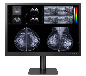
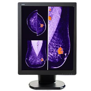
- Mammography- Mammograms are a type of x-ray image of the breasts. They screen for early evidence of breast cancer, sometimes identifying lumps years before they are palpable. A radiologist can use Digital mammography, which requires a much lower radiation dose to produce high-quality images of breast tissue to identify and diagnose cancer nodules that older systems can’t detect.
- Bone Density Scan- This procedure, also known as “bone mineral density testing,” uses x-ray equipment to measure the amount of bone minerals and calcium per square centimeter. Typically, this scan is conducted on the hip, spine, or forearm. This scan can determine whether a patient has osteoporosis (a condition where the bones are fragile and susceptible to fractures). Bone density scans are recommended for patients with the following risk factors: a recent fracture, height loss of more than an inch and a half, decreased hormone levels, long-term steroid use, or anti-rejection medication due to organ or tissue transplant.
- Arthrogram- Also known as “arthrography,” arthrograms consist of various images obtained using x-ray, fluoroscopy, CT scans, or an MRI specifically of an individual’s joints. An arthrogram is one of many types of medical imaging used to diagnose joint problems. To do this, a radiologist will inject your joint with a contrast dye that coats your joint structures and lining, allowing the physician to easily evaluate joint function. An arthrogram might capture detail that other types of imaging may not detect.
- Myelogram- During a myelogram, a technologist injects contrast dye into the spinal cord space. While this dye moves through the spinal spaces, fluoroscopy is used to examine the spinal cord, tissue, and surrounding nerves for any abnormalities, like tumors, infection, and inflammation.
- Nuclear Medicine – During this type of medical imaging, radioactive tracers are injected into a vein to provide images of the internal organs and structures. This gives physicians the opportunity to diagnose some types of cancers, gastrointestinal issues, or endocrine disorders. It can be used with a bone scan, thyroid scan, thallium cardiac stress, or positron emission tomography (PET scan).
Do you have imaging needs?

Double Black Imaging is dedicated to building long-term customer relationships by providing the world’s finest imaging components for diagnostic imaging at competitive prices. We are committed to supporting our products with exceptional customer service. Contact us today!