Human-Computer Interaction in Radiology & Reporting: A Look at Three Applications
Why Human-Computer Interaction in Radiology?
Focus on Human-Computer Interaction (HCI) in the field of radiology has increased significantly over the years. It is now common to use the terms HCI radiology and HCI imaging to refer to the study of radiology as it pertains to human-computer interaction.
The emphasis on HCI radiology, or HCI imaging, has changed the way we look at equipment purchases, report writing, and E-Learning.
The influence of HCI considerations not only has had a major impact on these areas to date but could continue to influence these and other considerations for the field of radiology in the future.
HCI Radiology and Equipment Purchases
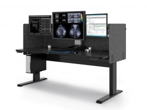
Jogia, et.al. (2021)1 reported a human-computer interaction study to consider HCI factors when purchasing X-ray equipment. While they looked at ergonomic factors such as force to operate machines, the primary emphasis was on the HCI between Medical Radiation Technologists (MRTs) and the control room console.
They focused on the tasks involved in operating the control room console as these are the actions that make up the HCI. Further, they looked at actions at the console that could result in wasted images and which aspects of the design might reduce repeated and wasting (rejecting) X-ray images.
A primary focus of the assessment involved heuristic evaluation. This allowed them to judge the usability without speaking to the users.
They identified three sources of the task workload for HCI activities: mental demand, frustration and performance.
Consistency and Standards, User Control and Freedom, Error Recovery Problems, Help and Documentation, and Error Prevention and Memory were all areas of errors when using the console.
They summarized the following questions:
Should HCI factors be included in a purchasing guide?
Can HCI factors be identified using HCI tools while observing MRTs working with the equipment?
Can we determine the most relevant method and tool for evaluating HCI between MRTs and the X-ray equipment?
Jogia, et. al. (2021) called for additional human-computer interaction studies employing the use of heuristic evaluation that focuses on factors that can be easily identified and tested during the purchasing process. They suggested that these HCI factors could be instrumental in planning and designing an X-ray suite.
HCI Imaging and Reporting
Ganapathi, et.al. (In print)2 studied HCI for the development of a structured reporting process that incorporated eye-gaze and speech signals. The primary objective was to generate data to create an HCI to automate generation of a structured radiology report that captures key image findings and a radiologist’s subsequent spoken descriptions of those findings.
They used an eye-gaze tracking device to establish an eye-gaze-voice span parameter which they defined as a unit of time from when the radiologist fixates on an image to the start of speech.
They looked at factors such as anatomy imaged, image modality, image resolution and radiologist interpretation.
They concluded that because the individual radiologist was the key to designing a system of automated reporting that utilizes eye gaze, a system based on these parameters would need to be customized to the individual radiologist.
The order of items targeted did not produce significant variability, nor did the anatomy being imaged, image resolution, and modality, so customization is not needed for these items.
They suggest a “next step would be to use the gaze and speech signals together to identify diagnostic regions-of-interest from the gaze signals and corresponding speech content to incorporate key image findings into a structured report.”
The authors believe that a system using eye gaze – voice span could improve structured acceptance and implementation.
The article concludes with the suggestion that the best success with the system could be achieved through training the radiologist to better use the HCI.
HCI Radiology and E-Learning
Den Harder, et.al. (2016)3 looked at how learning outcomes and perspectives of medical students and teachers were influenced using image interaction in radiology e-learning programs.
They defined e-learning as us of electronic media, including audio, digital images, and web-based learning for educational purposes.
Advantages of e-learning include flexibility in when and where a student can learn, the types of materials that can be used (animation, video, interactive programs), and the ability to increase class sizes.
Two primary forms of HCI that are of benefit to radiology in e-learning are:
Navigation or scrolling through a stack of images in different planes)
Manipulation (adjusting contrast setting, rotating 3D models)
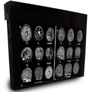
In the past, Computed tomography (CT) and magnetic resonance imaging (MRI) in hospitals were viewed by physicians as single images printed next to each other. In radiology, they are viewed as a stack of images (volumetric image). Now that images are digitalized and used in Picture Archiving Systems (PACS), the volumetric images are available to all in-house doctors.
This highlights the importance of medical students learning to interpret radiological images and understanding the relationship between the anatomical and pathological structures.
This article suggests that a “first step can be the use of videos of volumetric image stacks. However, ultimately, human-computer interaction with images should be possible since this is more representative to clinical practice and more reliable than tests with 2D CT images.”
They indicate that allowing for image manipulation is a key method e-learning can improve radiology education.
This was supported by the finding that tests that used stack images were more reliable and more representative of clinical practice compared to tests that used 2D images.
Now that in-hospital doctors have stacks of images available for review, and the study found that stack image and 2-D image review require different cognitive processes, the argument is made that medical students could benefit from similar exposure.
The study also noted that scores using stack viewing and image manipulation correlated with scores on human cadaver anatomy tests. This same correlation did not occur for tests without stack viewing.
The authors suggest that radiology anatomy courses should use radiological images with stack viewing and other image manipulation tools.
They concluded that the use of image interaction in e-learning could be beneficial for medical students.
About Double Black Imaging
 Double Black Imaging makes radiology imaging and reporting easier and less time consuming for you. Implementation of new information about HCI is just one way we hope to accomplish that.
We are here to help you solve your imaging issues and improve your process in your radiology department. Give us a call at (877) 852-2870, email us at sales@doubleblackimaging.com, or go here to submit a request. Let us know how we can help you use the latest knowledge to make your department work smarter.
References:
- Jogia, A; Brunet, J-P; Ramos, D; Lintack, J; Di Raimo, L; Sharpe, M; Rowr, K; Paul, N; Lall, D; Cheadle, S; Smith, J; Macdonald, R; Plastino, J. Human Computer Interaction (HCI) in General Radiography: A Case Study to Consider HCI Factors When Purchasing X-ray Equipment (2021). Proceedings of the 21st Congress of the International Ergonomics Association (IEA 2021), IV.
- Ganapathi, T; Vining, D; Bassett, R; Garg, N; and Markey, M. A Human Computer Interaction Solution for Radiology Reporting: Evaluation of the Factors of Variation. Journal Preprint.
- den Harder, A.M., Frijlingh, M., Ravesloot, C.J. et al.The Importance of Human–Computer Interaction in Radiology E-learning. J Digit Imaging 29, 195–205 (2016).
