Category: Uncategorized

30 Jan 2023

The Importance of Ergonomic Workstations in Radiology

Progress in medical science can be painful.  It’s no joke.  Technology such as PACS and digital imaging can cause repetitive strain injuries for radiologists.  And that’s just the beginning.

Radiology workstations designed ergonomically can help decrease the frequency and severity of repetitive strain injury.  Use of ergonomic principles can vary from simple changes to equipment that changes the way radiologists work and eliminates the cause of many injuries.

You’ve heard the term for several years now.  But what exactly is an ergonomic workstation, and how does it make things better?

Let’s start with the basics.

What is an ergonomic approach?

Almost every activity involved in a radiologist’s job can lead to some type of repetitive injury.

As a radiologist today, your job includes:

  • Viewing high contrast images in a dark environment multiple hours per day
  • Utilizing speech mics with Speech recognition software
  • In certain cases, performing procedures
  • Reporting test results
  • Preparation of lectures and presentations
  • More commonly, these tasks are being performed at home or from teleradiology workstations.

This is just a brief list of activities performed requiring repetitive motions.

Ergonomic workstations are designed to minimize the need for and impact of repetitive motions such as standing, bending, twisting, and leaning in performing your job.  Once these stressors are minimized, you can be more comfortable and productive when working.

What does an ergonomic workstation look like?

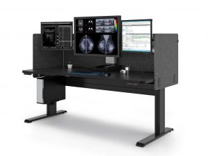
Ergonomic equipment for workstations varies by profession.  As it relates to radiology workstations, ergonomic equipment includes:

  • Desks/tables that have:
    • Sit to stand adjustment capability
    • Ability to change monitor height independent of worksurface
    • Ability to easily and quickly change focal depth of monitors
    • Large usable worksurface to provide space for radiologist’s equipment: multiple keyboards and mice, voice recognition mic, trackball, etc. . . Ideally a single surface to maximize useable space in a given footprint
    • Ability to be quickly and easily adjusted/set up for each radiologist using the station
    • Dimmable back lighting to reduce eye strain
    • Electric sit to stand workstations/desks
  • Monitors that:
    • Meet ACR and FDA standards for brightness and resolution, ensuring images are DICOM Calibrated for proper diagnosis
    • Include productivity tools with the ability to dim worklist panels, limiting ambient light causing eye strain.  Cursor wrap functions eliminating continuous scrolling over the worksurface
    • Maximize viewing clarity from various angles
    • Reduce stress by offering adjustable height and angle
  • Chairs that provide
    • Vertebral support
    • Head support

What should you consider before implementing an ergonomic approach at your radiology practice?

Implementing an ergonomic approach is more than buying equipment that has a list of features.  It is important to see how specific features impact your patient care, medical staff, and overall satisfaction of the team.

A comprehensive ergonomic workflow rollout address every aspect of the practice and includes proper training on the use of the equipment as well as how can prevent injuries.

Technicians in radiology can benefit from an ergonomic approach as well.  Important areas to consider for these team members includes:

  • Manipulation of equipment necessary for performing procedures
  • Transportation of the equipment
  • Sit/stand desks for MRI and CT technicians
  • Positioning of patients for completion of the procedure
  • Ergonomic seating

How Does an Ergonomic Workstation Help Your Practice?

Ergonomics offers more than physical benefits. It results in increased productivity and efficiency, increased accuracy of reporting, a comfortable work environment, improved employee retention, and improved patient care.

The Association for Talent Development1 indicated that there are several factors that are improved when you focus on health and wellness:

  • absenteeism
  • retention and turnover
  • productivity
  • employee engagement

The costs associated with these factors is not small.

When an employee is injured, employers pay for more than the worker’s medical treatment. In its “NSC Injury Facts 2017,” the National Safety Council (NSC) calculates the average cost of an injury at $31,0002.

Radiation therapists appear to be at considerable risk for work-related musculoskeletal injuries, which corresponds with findings of studies on health care employees. They are reported to have a high prevalence of injuries (76%)3.  In fact, 58% of radiologists report symptoms of repetitive stress injury (many spend over 8 hours per day at their desks).

The most common site-specific injury is the lumbar back, followed by the neck and shoulders3. Other injuries can include repetitive stress injuries, visual problems, and headaches.

Productivity and Efficiency

When an ergonomic approach is used, staff can avoid bending, twisting, and awkward positions that cause fatigue, soreness, and lost time, as well as decreased comfort as the workday progresses.

Employees can be happier and have more energy when they can be comfortable when they work.  It’s easy to see that more energy put into helping patients results in patients who are happier with the care they receive.

Accurate Data Entry: A Surprising Benefit

Initially you might not consider that an ergonomic approach to Radiology workstations could impact the accuracy of data entry.  However, consider that you would have a higher level of fatigue throughout the day when you have a high level of stress on your body. Errors are more likely due to the higher level of stress and fatigue that results in decreased focus.

Ergonomic equipment increases safety and comfort throughout the day, meaning that employees are safer, more comfortable, and less fatigued.

A Comfortable Radiology Workstation adds Value and Savings

When care providers can adjust table height, view records through an optimally sized and correctly positioned monitor, and move easily around the facility/unit, they are more comfortable and experience less fatigue. This means that strain on backs, wrists, and necks is drastically reduced, or eliminated.  Ergonomic workstations designed to the unique demands of a job helps the radiology team do their job effectively and efficiently in comfort.

A ROI Comes in Many Forms

By now you realize that implementing an ergonomic approach requires investment of time and money.

Considering the costs of worker’s compensation, rehabilitation, and training of new radiologists, it is easy to see how you can quickly realize a return on your investment.

The biggest savings are seen in:

  • Less time off for injury
  • Fewer staff injuries and worker’s compensation claims
  • Greater accuracy in reporting and patient care
  • Healthier and happier staff with less turnover

When preventable injuries, burnout, and staff error are avoided, money typically spent on these items can now be spent where it is needed most.

Improved Patient Care is the Ultimate Reward

Imagine the improved patient care and improved patient satisfaction when:

  • Procedures are completed once because the staff could effectively and efficiently do it correctly the first time
  • Appropriate care is provided because errors are not made that might be related to fatigue or limited focus
  • Patients are more confident in the services they receive because staff treating them are more positive and energetic when providing care

These are just a few ways that an ergonomic workstation can improve patient care.

Why You Should Work with Double Black Imaging for Your Radiology Workstations

At Double Black Imaging, we don’t just sell you equipment to meet a standard ergonomic need.  We complete an onsite radiology workstation survey to assess what ergonomic equipment will be best for you and your radiologists.

We make specific recommendations for each surveyed workspace.  Our recommendations for your specific needs are based on our insider knowledge of PACS, mammography/tomography, clinical and surgical applications.

Contact us here, email us, or call us at (877) 852-2870, to arrange your Onsite Radiology Equipment Survey and determine what workstation will be best for you and your radiologists.


References:

  1. ATD Releases 2016 State of the Industry Report | ATD, January 9, 2023
  2. Employee Injuries: What Is the Real Cost to Employers? (workplacetesting.com), January 9, 2023
  3. Prevalence of Musculoskeletal Work-related Injuries Among Radiation Therapists – PubMed (nih.gov), January 9, 2023 Hanania AN, Cook A, Threadgill MP, Conway SH, Ludwig M. Prevalence of Musculoskeletal Work-related Injuries Among Radiation Therapists. Radiol Technol. 2020 May;91(5):414-421. PMID: 32381659.
17 Nov 2022

Benefits Of Mobile Radiology Imaging

Mobile Imaging services mean that certified technologists and radiologists travel to a medical facility, residence, or place of business with any equipment needed to complete a procedure and provide those services at that location.  It not only includes portable X-Ray imaging but also ultrasound and EKG.

Mobile services are increasing in popularity because of changes in the way facilities administer healthcare and assess improvement.  Because mobile imaging services can provide faster, more cost-effective, and more convenient services compared to third-party facilities, they are being used more often.


Patient Benefits of Providing Mobile Imaging


Typically, patients would expect to receive radiology services such as X-Ray, ultrasound, or EKG by traveling to a third-party imaging center or an in-house imaging department, or as an inpatient in a hospital traveling within the facility to the imaging department.

Faster and Cheaper Services

Besides being able to schedule an appointment faster than with traditional service providers, mobile imaging results can be communicated faster to the ordering physician.   This is possible because mobile imaging services are available 24 hours per day and often have 24-hour turnaround times. Another factor that makes mobile services faster is that the scans are available via online portals such as PACS within minutes.

Mobile radiology services can be faster and cheaper than those available through traditional models at the sites discussed above. Reduced expense is achieved by having shorter response times and greater flexibility with mobile imaging. It is also cheaper for the patient because mobile imaging does not require patient travel to a facility for services.

Patient Care Benefits

Patient health is risked when the care team needs to interrupt the peace and quiet of the room to prepare the patient for transportation, transfer them to a different bed for travel, and/or disconnect treatment and transfer it to the transportation device.  When patients are already very ill, transportation increases risks.

Disruption of a tranquil environment presents health risks whether you are talking about the patient being transported or the patient in the room and who is also having their tranquility disrupted.  A quiet environment can help patients heal faster because it helps the patient sleep better.  When a patient is in a quiet environment, they experience fewer spikes in heart rate, decreased cortisol output, lower blood pressure, and reduced anxiety.

It should be noted that fewer staff need to enter the room and work with the patient when the imaging service comes to their room.  Because fewer people are entering the room and working with the patient at the same time, this results in less disruption to other patients in the room.


Avoiding Health Risks

Patients referred for imaging services while hospitalized face several risks to their health and safety during transportation to the Imaging department.  While being transported, the patient could be exposed to several health and safety risks, including:

  • infections (air-born acquired illnesses, infections from wounds, contact-born pathogens)
  • respiratory problems (ventilator, tracheostomy, and breathing assistance)
  • traumatic injury (falls, lacerations, hematomas)
  • medical complications (glucose regulation or acid-based homeostasis)
  • death

Avoiding Hospitalizations

In the past, a large number of hospital admissions of patients from nursing homes and skilled nursing facilities were related to falls, respiratory infections, and digestive system inabilities.  Being able to assess these patients at their residence can decrease the need for hospitalization.  Now many patients can be diagnosed, and a treatment plan can be identified without the need for hospital admission.


Facility/Staff Benefits


Decreased Costs

Mobile radiology imaging can be integrated into current services without extensive overhead costs.

From the facility’s perspective, it requires far fewer personnel to provide a mobile scan than one that is required to prep and transport a patient, especially if they are very ill and have multiple devices that must travel with them.

Busy third-party imaging facilities and hospitals suffer financial losses when many patients cancel on the same day due to inclement weather.  With mobile services, the patients stay where they are, and the imaging services come to them.

Mobile imaging services can be incorporated into your current facility and service delivery model without adding space, equipment, or training.

Faster Results and Better Patient Treatment

Mobile imaging services can provide support to in-house imaging departments.  This assistance provides a quicker response to patients when the in-house service is not able to meet the demand of both in and outpatients.

Another variable that makes mobile imaging faster and less expensive is the turnaround time for disinfection.  Most mobile units can be disinfected and ready to use again in as few as 5 minutes compared to the 45 minutes that is usually required for a radiology suite.

Greater Medical Team Satisfaction

When imaging services come to the patient, the medical team experiences less stress.  Since fewer professionals are required to assist the imaging team, the medical team on the nursing unit is freed up to help other patients. Therefore, there can be fewer simultaneous demands on the team member’s time and energy.


Types of Imaging Available in a Mobile Setting


Currently, mobile imaging services include X-rays, EKGs, and ultrasound.  It is reasonable to expect that this list of services will increase in the future.

X-Ray

X-Ray procedures are performed for a variety of reasons.  They are used to detect gallstones and kidney stones, injury to bones, bone deformities, pneumonia or bronchitis, and even aspiration.  X-rays can also be used to verify bone healing or the placement of medical devices such as catheters, implantable pumps, or feeding tubes.

EKG

EKGs performed by a mobile service are used for the same purposes as those performed in-house.  They are used to identify heart attack, pericarditis, or angina as a cause of chest pain.  Also, it can identify the cause of symptoms of heart disease like shortness of breath, dizziness, fainting or rapid heartbeat, irregular heartbeats, or palpitations.  EKGs can also monitor whether medications are improving a patient’s condition or whether they are causing side effects that impact cardiac functioning.  Finally, EKGs can assess heart health in the presence of concomitant conditions such as high blood pressure, high cholesterol, diabetes, heart disease, or smoking.

Ultrasound

Ultrasound performed by a mobile radiology imaging service can detect various medical conditions, including abdominal aneurysms, arterial occlusion or blood clots, carotid occlusive disease, carotid artery disease, renal vascular disease, hypertension, and early signs of kidney failure.


Double Black Imaging Can Help With your Imaging Needs


Double Black Imaging has been working to make imaging more efficient since 2002.  We focus on keeping in step with new technology and trends in the industry.  It is also our passion to help you, our customers, stay on top of those trends.

We are known for our customer service and pride ourselves on providing the best in the industry.  Why?  Because we believe our job is to make your job easier.

One way to make work easier at your facility is to help you integrate mobile imaging services.

Are you ready to make that happen?  We are here to help you make it effortless, effective, and efficient.

Contact us here, email us at sales@doubleblackimaging.com or call us at (877) 852-2870.  We look forward to helping you succeed!


25 Mar 2022

No Surprises Act in Radiology

No Surprises Act in Radiology: A Summary of Key Factors

What is the No Surprises Act in Radiology?


When treated by facilities or physicians outside their insurance network  patients could receive large, unexpected bills. This is known as surprise billing.

Simply stated, these providers do not participate in an insurance plan and typically bill and are reimbursed at a different rate.

The No Surprises Act is a federal law that limits the financial obligations of patients to out-of-network providers. This applies to emergency and non-emergency situations.

No Surprises Act billing rules state that a patient’s out-of-pocket expenses should be the same for out-of-network providers as it is for providers in-network. Depending on the patient’s insurance plan, this will be the co-payment, co-insurance, or deductible that is normally owed to in-network providers.


When was it voted on?


In December 2020, Congress signed the Consolidated Appropriations Act.  The No Surprise Act is part of that legislation.


When did it go into place?


The No Surprises Act effective date was January 1, 2022, so you are now expected to follow the law.


What has changed because of this act?


If your state has already enacted no surprise billing legislation, the No Surprises Act will not supersede it.

For states that have not yet enacted a surprise billing law, the No Surprises Act will result in several changes.

For non-emergency care, you will need to obtain consent prior to providing services.

Secondly, the patient cannot be billed directly.  You will need to bill and negotiate reimbursement through the insurance provider.

Once you have received payment, you have 30 days to negotiate or appeal the amount.

Finally, you will need to use the established system to negotiate and/or request arbitration of reimbursement rates.


Services Covered


If a patient requires emergency care, you should provide services until they are stabilized.  As an out-of-network facility, you must then address the issue with the patient to obtain consent to be transferred to an in-network facility or physician.

The patient can also consent to continue receiving out-of-network services from you.

Patients who sign consent waivers will then be responsible for the balance of uncovered expenses.

In a non-emergency situation, if your facility has in-network providers, you are permitted to request a consent waiver.  This allows you as the provider to balance bill the patient.

In these non-emergency situations, it is important to note that you need to give the patient a written consent waiver 72 hours prior to the procedure/medical service.

You need to include the following in the consent waiver:

  1. A declaration that you are not in the patient’s insurance network
  2. An estimate of the cost of your services that is as accurate as possible
  3. If your facility is in-network, a list of in-network providers at your facility
  4. A summary of any prior authorization or care management requirements that must be in place prior to the procedure/service
  5. A statement that the patient can opt to receive care from an in-network provider

When the non-emergency procedure will occur in less than 72 hours, you must provide the notice/consent waiver at the time the appointment is made.


Independent Dispute Resolution (IDR)


The No Surprises Act provides a system for negotiating reimbursement.  Please note, you only have 30 days to come to a resolution with the payer or to file a dispute if you cannot come to an agreement.

If you fail to reach an agreement with the payer, you or the payer have four days to activate the Independent Dispute Resolution (IDR) system.

Once the IDR is initiated, you and the payer will each submit an amount to an arbitrator, who will select one of those amounts as the settlement.

You cannot split the difference or request further negotiation.

Once decided, neither you nor the payer can repeat the process for the same service or procedure with each other for 90 days.

The losing party pays the cost of the arbitration process.

Over time, you will learn what level of payment is acceptable to the arbitrator, and this should limit the need for IDR appeals.


Impact on Radiologists


Radiologists and other ancillary service providers are not able to use consent waivers. This means you must abide by the in-network payment policies of the Act.

The No Surprises Act does not exempt patients from financial responsibility. You can expect them to pay what they would pay if you were in-network. This will be clearly communicated to you on the Explanation of Benefits (EOB) that the insurance company sends with their payment to your practice. You can bill the patient for this amount once you have accepted the payment or a payment has been negotiated. You can bill them for their co-payment, co-insurance, or deductible as indicated in their insurance plan.

Time and resources spent negotiating rates and engaging in arbitration, will impact finances for you and the payer. It is in your best interest to establish a system to minimize this impact.

One solution is to try to reach an agreement with out of network (OON) payers. This will eliminate potential disputes about reimbursement, special claim handling and costs of arbitration via IDR, so it will be beneficial to them to make this agreement with you.


Importance of developing an internal process


You will learn about the charges and reimbursement for OON patients as you settle disagreements with the payer.  However, handling charges for these patients will require a different system than what you use for in-network billing.

Be sure in-network vs. out-of-network status is clearly determined from the beginning for these patients so their reimbursement claims can be processed correctly.

Even though patients are out-of-network, you should submit your initial claim to the insurance company for your full fee.

Your system should tag accounts so that you can review payments when they are received.  This will avoid billing the patient for the balance rather than the allowed amounts.

You also want your staff to determine whether the payment is acceptable to your practice within the 30-day negotiation window.  This way, you don’t miss the opportunity to negotiate a rate or file a dispute.

Your staff can respond quickly and know if negotiation is necessary. If you take the time to create a list of reimbursement rates that you find acceptable for your procedures. Also, over time you will learn from the arbitrator what is an acceptable reimbursement level.

Claim processing can be more efficient if you create a fee schedule within your accounting system.


About Double Black Imaging


Learn More About No Surprises Act in Radiology

Since 2002, Double Black Imaging has brought high quality computing and imaging products to the market in response to changes in the market technology and medical care. We offer solutions that will help you implement these changes effectively and with minimal effort.

Contact us today at (877) 852-2870 or email us at sales@doubleblackimaging.com to learn more about how our products can help you comply with these new requirements without a negative impact on your budget.

23 Feb 2022

Human-Computer Interaction in Radiology & Reporting: A Look at Three Applications

Why HCI in Radiology?


Focus on Human-Computer Interaction (HCI) in the field of radiology has increased significantly over the years.  It is now common to use the terms HCI radiology and HCI imaging to refer to the study of radiology as it pertains to human-computer interaction.

The emphasis on HCI radiology, or HCI imaging, has changed the way we look at equipment purchases, report writing, and E-Learning.

The influence of HCI considerations not only has had a major impact on these areas to date but could continue to influence these and other considerations for the field of radiology in the future.


HCI Radiology and Equipment Purchases


Jogia, et.al. (2021)1 reported a case study to consider HCI factors when purchasing X-ray equipment.  While they looked at ergonomic factors such as force to operate machines, the primary emphasis was on the HCI between Medical Radiation Technologists (MRTs) and the control room console.

They focused on the tasks involved in operating the control room console as these are the actions that make up the HCI.  Further, they looked at actions at the console that could result in wasted images and which aspects of the design might reduce repeated and wasting (rejecting) X-ray images.

A primary focus of the assessment involved heuristic evaluation.  This allowed them to judge the usability without speaking to the users.

They identified three sources of the task workload for HCI activities:  mental demand, frustration and performance.

Consistency and Standards, User Control and Freedom, Error Recovery Problems, Help and Documentation, and Error Prevention and Memory were all areas of errors when using the console.

They summarized the following questions:

Should HCI factors be included in a purchasing guide?

Can HCI factors be identified using HCI tools while observing MRTs working with the equipment?

Can we determine the most relevant method and tool for evaluating HCI between MRTs and the X-ray equipment?

Jogia, et. al. (2021) called for additional studies employing the use of heuristic evaluation that focuses on factors that can be easily identified and tested during the purchasing process.  They suggested that these HCI factors could be instrumental in planning and designing an X-ray suite.


HCI Imaging and Reporting


Ganapathi, et.al. (In print)2 studied HCI for the development of a structured reporting process that incorporated eye-gaze and speech signals.  The primary objective was to generate data to create an HCI to automate generation of a structured radiology report that captures key image findings and a radiologist’s subsequent spoken descriptions of those findings.

They used an eye-gaze tracking device to establish an eye-gaze-voice span parameter which they defined as a unit of time from when the radiologist fixates on an image to the start of speech.

They looked at factors such as anatomy imaged, image modality, image resolution and radiologist interpretation.

They concluded that because the individual radiologist was the key to designing a system of automated reporting that utilizes eye gaze, a system based on these parameters would need to be customized to the individual radiologist.

The order of items targeted did not produce significant variability, nor did the anatomy being imaged, image resolution, and modality, so customization is not needed for these items.

They suggest a “next step would be to use the gaze and speech signals together to identify diagnostic regions-of-interest from the gaze signals and corresponding speech content to incorporate key image findings into a structured report.”

The authors believe that a system using eye gaze – voice span could improve structured acceptance and implementation.

The article concludes with the suggestion that the best success with the system could be achieved through training the radiologist to better use the HCI.


HCI Radiology and E-Learning


Den Harder, et.al. (2016)3 looked at how learning outcomes and perspectives of medical students and teachers were influenced using image interaction in radiology e-learning programs.

They defined e-learning as us of electronic media, including audio, digital images, and web-based learning for educational purposes.

Advantages of e-learning include flexibility in when and where a student can learn, the types of materials that can be used (animation, video, interactive programs), and the ability to increase class sizes.

Two primary forms of HCI that are of benefit to radiology in e-learning are:

Navigation or scrolling through a stack of images in different planes)

Manipulation (adjusting contrast setting, rotating 3D models)

In the past, Computed tomography (CT) and magnetic resonance imaging (MRI)  in hospitals were viewed by physicians as single images printed next to each other.  In radiology, they are viewed as a stack of images (volumetric image).  Now that images are digitalized and used in Picture Archiving Systems (PACS), the volumetric images are available to all in-house doctors.

This highlights the importance of medical students learning to interpret radiological images and understanding the relationship between the anatomical and pathological structures.

This article suggests that a “first step can be the use of videos of volumetric image stacks.  However, ultimately, human-computer interaction with images should be possible since this is more representative to clinical practice and more reliable than tests with 2D CT images.”

They indicate that allowing for image manipulation is a key method e-learning can improve radiology education.

This was supported by the finding that tests that used stack images were more reliable and more representative of clinical practice compared to tests that used 2D images.

Now that in-hospital doctors have stacks of images available for review, and the study found that stack image and 2-D image review require different cognitive processes, the argument is made that medical students could benefit from similar exposure.

The study also noted that scores using stack viewing and image manipulation correlated with scores on human cadaver anatomy tests.  This same correlation did not occur for tests without stack viewing.

The authors suggest that radiology anatomy courses should use radiological images with stack viewing and other image manipulation tools.

They concluded that the use of image interaction in e-learning could be beneficial for medical students.


About Double Black Imaging


Radiology Monitor Provider and RetailerDouble Black Imaging makes radiology imaging and reporting easier and less time consuming for you.  Implementation of new information about HCI is just one way we hope to accomplish that.

We are here to help you solve your imaging issues and improve your process in your radiology department.  Give us a call at (877) 852-2870, email us at sales@doubleblackimaging.com, or go here to submit a request.  Let us know how we can help you use the latest knowledge to make your department work smarter.


References:

  1. Jogia, A; Brunet, J-P; Ramos, D; Lintack, J; Di Raimo, L; Sharpe, M; Rowr, K; Paul, N; Lall, D; Cheadle, S; Smith, J; Macdonald, R; Plastino, J.  Human Computer Interaction (HCI) in General Radiography:  A Case Study to Consider HCI Factors When Purchasing X-ray Equipment (2021).  Proceedings of the 21st Congress of the International Ergonomics Association (IEA 2021), IV.
  2. Ganapathi, T; Vining, D; Bassett, R; Garg, N; and Markey, M. A Human Computer Interaction Solution for Radiology Reporting:  Evaluation of the Factors of Variation.  Journal Preprint.
  3. den Harder, A.M., Frijlingh, M., Ravesloot, C.J. et al.The Importance of Human–Computer Interaction in Radiology E-learning. J Digit Imaging 29, 195–205 (2016).

26 Oct 2021

Ergonomics in Interventional Radiology

Professionals in interventional radiology perform medical imaging during various minimally-invasive procedures such as X-ray fluoroscopy, computed tomography, magnetic resonance imaging, or ultrasounds. These imaging tests can help diagnose, cure, or alleviate symptoms of vascular disease, stroke, cancer, and much more.

Although times may vary depending on each individual case, interventional radiology procedures usually take approximately four hours. During any invasive procedure, the patient’s safety is the top priority, which is why a lot of interventional radiologists neglect basic ergonomic needs and ultimately, their health during procedures. This means interventional radiologists are regularly exposed to radiation, and lack of ergonomic equipment can lead to work-related musculoskeletal disorders in the long term.

In fact, in interventional radiology, the use of x-ray aprons, in association with awkward postures and non-ergonomic working conditions, might increase the likelihood of musculoskeletal disorders. The prevalence of neck and back pain at least once a week ranges from 50% to 60% for those who use lead aprons frequently. Additionally, not having the right equipment to work with, will likely impact their livelihoods, quality of life, and productivity in various ways.

Even though interventional radiology often overlaps with other specialties such as cardiology, vascular surgery, orthopedic surgery, and urology among others, implementing unique ergonomic considerations is key to ensure interventional radiologists’ safety in their place of work. Having an interventional radiology suite with ergonomic equipment is crucial to the prevention of musculoskeletal disorders (MSDs) and injury. By including ergonomic interventional radiology equipment in your facility, you are investing in the health, wellbeing, and productivity of interventional radiologists.


Here are a few things to consider to maximize your investment and efficiently plan for a new interventional radiology suite:


What makes up an efficient interventional radiology suite?


To optimize interventional radiologists’ performance, as well as improve their wellbeing while meeting patients’ needs, there are various crucial aspects to consider when planning for your suite. One of the key things to keep in mind is the types of procedures the equipment will be used for. There are often various professionals that are part of a procedure, such as nurses, surgeons, anesthesiologists, and radiologists. Understanding each professional’s role and the input will ensure you plan your investment accordingly.

Each area of expertise requires its own set of equipment and standards to follow during the installation. The Society of Interventional Radiology published “Resource and Environment Recommended Standards for the IR” where it details recommendations for the interventional radiology surgical suite.

Furthermore, The Society of interventional Radiology recommends: “The preparatory and recovery areas must be located in a setting with adequate electrical, oxygen, suction, anesthesia, and emergency services. There must be appropriate temperature and humidity control, air exchange and ventilation, lighting, computer terminals, PACS access, and monitoring equipment. Access to basic and advanced resuscitation equipment as well as necessary medications and fluids and adequate support area for supplies, nutritional support, ice machines, blanket warmers, and nursing space is also required.”

How is interventional radiology equipment different?


The main focus when purchasing new equipment should be to make interventional radiologists’ workflow more ergonomic and make patient care more personalized. The following is a list of essential equipment that is required in an interventional radiology suite:


  • Imaging equipment:

Limiting radiation exposure is key for IRs. While lead aprons are commonly used they can weigh up to 15 pounds and increase pressure in the lumbar or cervical discs. Instead, today’s mobile technology like CT scanners can provide improved quality without high radiation exposure. Because interventional radiology is used during invasive procedures, it’s key for both radiologists and surgeons to clearly see what they’re working on. Using surgical displays can ensure any issues that come up are addressed immediately. Ultimately, they make any procedure safer while ensuring issues can be observed in real-time during crucial operations.

  • Ultrasound equipment:

Awkward positions are often adopted by professionals performing ultrasounds. Upper extremities are usually sustained in forceful gripping for long periods of time and are often conducive to work-related MSDs. In fact, ​​studies found that the average scan can lead to back, neck, and shoulder pain. To avoid the risk of MSDs, IRs should vary their exam postures throughout their working day.
Enter ergonomic workstations. Every radiologist needs the easy adjustability that an ergonomic desk setup will provide to stay productive. Ergonomic workstations that include adjustable heights, give IRs the possibility to switch their position, limiting the risk of postural and visual fatigue. Whether IRs are working sitting down or standing up, monitors should be placed just below eye level so that the neck muscles are relaxed at a downward viewing angle.


  • Medical grade computer monitors:

Today, these crucial devices are revolutionizing the medical industry with their ability to provide faster, cost-effective, and convenient imaging services in contrast with traditional imaging departments in hospitals and third-party facilities. Using a large screen and a broadcast video system may allow an ergonomic multimodal visualization that ensures IRs, surgeons, and any other key personnel attending a procedure has a clear picture.


Why interventional radiology equipment is key to improving your radiology teams efficiency


Work-related MSDs don’t only cause pain and discomfort during procedures, but affect IRs during their time off work. This can lead to burnout, a response to stress that involves both physical and emotional exhaustion and cognitive weariness. The risk with healthcare professionals having burnout is that ultimately it can affect their cardiovascular health and cause depression. However, promoting the wellness of IRs can help prevent this.

Interventional radiology suite design was created to make a safer, more efficient, and productive work environment for IRs. By investing in an IR suite, you can further optimize the performance and well-being of IRs while significantly reducing the risk of MSDs in your healthcare facility.


Get started with an ​​interventional radiology suite that will make your radiology practice more productive


Ergonomics In RadiologyWhen it comes to making an important investment in your healthcare facility, you want to make sure you do so through a reliable company that has a thorough understanding of your needs. With over 30 years of experience in the high-performance display industry, Double Black Imaging provides ways to make imaging more efficient. Dedicated to building long-term customer relationships, they are committed to supporting quality products with exceptional customer service.

Double Black Imaging offers the latest mobile medical imaging technology as well as ergonomic workstations at competitive prices and can walk you through how to implement it in your healthcare organization. Contact Double Black Imaging for help with making the most of your purchase, or take a look at our latest ergonomic workstations.

17 Sep 2020

Radiologist Burnout

Why Does It Happen and How Can You Prevent It?

Although most physicians choose to become a radiologist because they see it as a meaningful career, burnout in radiology is at an all-time high compared to other medical specialties. In the 2018 Medscape National Physician Burnout and Depression Report, radiology was ranked as the seventh highest specialty for burnout among physicians. This is compared to its 20th highest ranking in 2017. The 2018 report found that 42% of 15,543 physicians surveyed reported burnout, with 45% of these physicians being radiologists.
Radiologist Burnout

What is Radiologist Burnout?

Burnout is characterized by feelings of being emotionally depleted, and a sense that one is accomplishing less and experiencing less satisfaction in one’s role. It is often accompanied by depersonalization where the physician feels disconnected from his or her work and peers, and where patients are no longer treated with empathy as individuals in pain or crisis due to indifference.

Impact of Radiologist Burnout

Burnout in radiology can lead to patient errors and omissions as well as other adverse events. In addition, there is the potential for lost productivity, and therefore lost income, when a physician is not able to function optimally or is absent for physical or mental health reasons. These are high costs to pay for burnout, not to mention the toll it takes on a physician’s mental and physical well-being.

Other results of burnout include:

  • • Physician attrition from the profession
  • • Early retirement resulting in more physician shortages
  • • Increasing health care costs
  • • Increased risk of substance abuse
  • • Increased risk of suicide


Early Warning Signs of Radiologist Burnout

Being able to recognize when you or one of your colleagues is beginning to show signs of burnout is important so it can be caught and addressed as soon as possible. Some early warning signs include:

      1. Complete exhaustion

        Exhaustion may be a feeling of emotional, mental, and/or physical fatigue. Signs of exhaustion or fatigue include:

        • • Difficulty waking up or getting out of bed in the morning
        • • Inner resistance to going to work
        • • Feeling of having no energy
        • • Irritability
        • • No desire to socialize after work with friends or family or participate in activities and hobbies
        • • Lack of desire to engage in personal wellness activities such as exercise and cooking at home


    1. Emotional detachment

      One way of coping with emotional fatigue and burnout is by detaching emotionally from patients. This emotional detachment can look different for different people, but may include cynicism towards patients and a more critical attitude when dealing with colleagues and staff. For others, sarcasm is used to emotionally detach from patients and deal with frustration.

    2. Work is dominating your life

      If you feel like your work is taking over your life in an unhealthy way, it probably is. If you find yourself constantly thinking about work even when you are trying to do non-work-related things, this is a warning sign. Because of the exhaustion that accompanies burnout, you may find your personal and social relationships suffering as well. And if you have no desire or ability to do anything but work, this is a sign you are in trouble.

    3. Feel like work is pointless and/or without a purpose

       Having bad days and feeling frustrated at times is part of any job. However, if you find yourself questioning why you are doing the work you are doing, or feel what you do is not making a difference, you are likely experiencing burnout from your job.

    4. Mistakes are more frequent

       When you are exhausted, frustrated, and emotionally detached from your patients and those you work with, mistakes are more likely to happen. If you find yourself making mistakes more often than usual, or making errors you would not usually make, it may be time to step back and reevaluate what is happening since this can be a sign of burnout.


Steps to Address Radiologist Burnout

Healthcare facilities and organizations are able to play an important role in helping reduce burnout and increase radiologist job satisfaction. Some strategies include:

  • • Recruit and assign radiologists to work in areas they prefer, enjoy, and are skilled and certified in.
  • • Avoid assigning work to radiologists who lack the certification or skill to perform the work competently and safely.
  • • Identify ways to reduce time radiologists spend on administrative activities by hiring assistants and considering virtual scribes to perform these activities instead.
  • • Support and encourage strong teams of healthcare professionals that interact regularly to assess workflow efficiency and work-related needs and difficulties of team members.
  • • Learn how to assess and recognize ongoing frustration and impending burnout in healthcare professionals and identify caring ways to offer assistance.


How to Avoid Burnout as a Radiologist

Some important steps you can take to avoid burnout in radiology include:

  • • Avoid working in isolation when possible
  • • Work with your peers and staff to create a positive work environment that supports one another
  • • Take frequent breaks from your workstation
  • • Get outside or away from your work area entirely during breaks
  • • Consider setting up an ergonomic sit/stand combination workstation
  • • Identify and engage in regular self-care habits that help you recharge physically, mentally, and emotionally
  • • Schedule days off regularly and take personal or vacation time off work
  • • Revisit regularly why you chose to become a radiologist and all the people you are helping (and have helped) with your expertise


References:

https://www.diagnosticimaging.com/view/stop-burnout-radiology-it-starts

https://www.radiologytoday.net/archive/rt0419p24.shtml

https://www.ncbi.nlm.nih.gov/pmc/articles/PMC6530597/

https://www.radiologybusiness.com/topics/leadership/radiologist-burnout-are-we-done-yet

https://www.mededwebs.com/blog/meded-manager/symptoms-of-physician-burnout-know-the-warning-signs

https://www.physicianspractice.com/view/five-signs-physician-burnout

https://www.gehealthcare.com/feature-article/decreasing-radiologist-burnout

17 Aug 2020

Imperfect Images and Discrepancies in Radiology

While the technology in medical imaging is constantly improving, what seems to be constant is the imperfect interpretation of images in radiology. According to a report that was released by the Institute of Medicine (IOM) in 1999, it is estimated that between 44,000 and 98,000 US citizens lose their lives each year due to preventable diagnostic errors. In a more recent study, these numbers have risen to 251,000 deaths. Furthermore, a third of the deaths stem from medical errors, which are heavily impacted by the diagnostics of practicing radiologists. (Waite, 2017)

Errors in the field of radiology image reading are not new according to the pioneer radiologist Leo Henry Garland (1903–1966). He found that even well skilled radiologists make mistakes in interpreting the images. Followed by other studies since Garland, the aim of the scientific studies has been geared towards understanding the basis of such errors and finding strategies to eliminate them. The categories of errors in radiology have been identified as perceptual and interpretive. That being said, perceptual errors are the most common. (Bruno, 2015)
nbsp;

Radiology Education

 
Before discussing the reasons for these errors, it is important to consider the preparation for the profession. Educating medical doctors in the field of radiology takes many years. Even then, mistakes are being made, which leads a person to wonder if there is a flaw in the educational system. This is according to Eric Postal, MD, who is the author of the article “Why Don’t Radiology Textbooks Have Imperfect Images?”, which is published on the Diagnostic Imaging website. He found that there may be a disconnect between the radiology textbooks and the actual medical imaging practices and diagnosis.

Eric Postal, MD was a medical student who found that textbooks did not prepare medical students for what to expect when working with real X-Ray images, let alone how to prevent discrepancies in radiology reading. Postal said that it was some time in his radiology residency that he realized something. This was that there were considerable differences between what he saw in textbooks and conferences, and what he saw in cases and reading-rooms.

What he found was that the textbook images were not the same as what a medical professional would see when working in a hospital. He found that the textbook images were perfect for learning purposes, but they did not prepare students for what to expect from real X-Ray images. For instance, “ultrasound pics suffered from shadowing by bowel-gas, or patient inability to position, suspend respiration, etc.,” he noted. What he noticed was that real X-Ray images were heavily distorted for many reasons, such as thorough shadowing by the bowel-gas. Consequently, medical students are bound to go through a period of adjustment when trying to analyze X-Rays they did not get prepared to read in medical school. (Postal, 2019)
nbsp;

Most Common Reasons for Radiology Imaging Errors

 
Errors are inevitably found in every branch of the scientific field, including radiology. Furthermore, the medical field of radiology is far from being exempt from this nature. Imperfect images that are placed before the practicing radiologists are subject to interpretations, making errors a likely outcome. Sometimes there may not be one clear answer but, instead, possibilities of answers. In such uncertainties, it is quite understandable that numerous amounts of errors do happen.[11]

Complexity of medical diagnosis puts the radiologist in a vulnerable position against making mistakes. It is interesting to note that errors made in clinical diagnosis are positively correlated with the errors made in radiological diagnosis. Thus, it is well worth our time to take a closer look at the errors that could be made and how to prevent these discrepancies in radiology. The following are articulations of how mistakes are easily made within the radiology sector.
  • Unclear images:
    Whether it is due to inadequate, old equipment or patients who fail to position themselves well enough for a clear image, radiologists struggle when reading images that are fuzzy, distorted, and unclear. This mistake alone is not due to the lack of the radiologist’s ability, but a result of the lack in quality of tools and resources. (Bruno, 2015)


  • Complexity of information:
    Images vary tremendously from one piece of equipment to another, especially when one machine is more advanced than the other. Comparing old images received from older equipment with the images received from newer equipment provide numerous challenges in reading and understanding what is going on in an individual’s body. With the advancing technology of imaging, radiologists are faced with learning to interpret new images at a fast pace. Sometimes reading images from newer equipment may take longer due to the lack of training on reading images that are produced from newer equipment. In cases of urgency for medical treatment decisions, late interpretation may impact patients tremendously. (Brady, 2012)


  • Fast paced work environment and fatigue:
    Work overload could be a common issue in radiology. When there are many images to interpret and very little time to interpret them, let alone write reports clear enough to present it to other healthcare professionals, it often results in the radiologist feeling great amounts of fatigue. In cases like this, the error margin increases due to lack of focus from the reader. (Bruno, 2015)


  • Distractions and Interruptions:
    Office related distractions and interruptions such as phone calls, e-mails, questions from other office staff, remote teleconferencing with other health specialists, just to name a few, all impact on the error margins negatively. (Bruno, 2015)


  • Satisfaction of the research:
    Detection of a focused abnormality and diagnosing it may cause the radiologist satisfaction of his/her work and terminate the interpretations prematurely. In cases like this, other underlying abnormalities that may or may not be related to the focused research, but they are easily missed and cause for unnecessary errors. (Bruno, 2015)


  • Under-reading:
    Abnormalities may be clear in the images, but the radiologist may overlook and miss seeing the abnormality. Reporting such errors without further detection could cause severe fatalities in patients’ lives. (Bruno, 2015)


  • Negligence:
    Medical professionals are not exempt from making mistakes in their career. However, most of these mistakes are either corrected after later detection or remain relevant to patients’ current state in health. When such mistakes exceed the expected error margin, it is considered negligence and may cause the radiologist to face legal disruptions. (Bruno, 2015)


  • Complacency:
    The images may be clear, and the radiologist may be able to identify the abnormality, but interpreting the image in order to find an underlying cause may not be as easy, which has caused many misdiagnoses. Sometimes this may be due to cognitive error, and the radiologist may interpret the reading differently than the real cause. (Bruno, 2015)


  • Lack of knowledge:
    Not all radiology readers are well prepared for their position, and sometimes they lack the necessary knowledge in order to effectively complete the task at hand. The abnormalities may be identified correctly on the images, but the importance of the diagnostic may be overlooked due to lack of knowledge or experience. (Bruno, 2015)


  • Poor Communication:
    In some cases, the abnormalities could be correctly identified, images clearly interpreted, and diagnosis is accurately made. Yet the written report either does not reflect the accurate information or it is not delivered to the physician of the patient due to poor communication channels between the two offices or (Bruno, 2015)


  • Other complications:
    Sometimes the abnormalities are not clearly present in the images for the radiologists to identify due to poor examination or poor technique. Sometimes, errors occur due to inaccurately diagnosed medical history of the patient which might lead the radiologist to inaccurate diagnosis. (Bruno, 2015)


  • Shortage of staff:
    Shortage of staff could cause the workload for every working individual to be overwhelming. As imaging techniques advance, and the study of images increases, the demand for the radiology staff that can interpret the images also increases.


  • Lack of adequate information:
    Lack of adequate information shared with the radiologist could cause errors in the diagnosis. Through these errors, it is proven that the use of teleradiology reporting from remote locations is not beneficial.  The more collaboration between the physician and the radiologist, the better and more accurate treatment decisions are made for the patients.


  • Location errors:
    Location errors in images occur when the objective of the photograph is not visible due to its location. Either the intended focus of the image is on the outside of the image or it is taken from an angle at which the abnormality is easily unidentifiable, because of an overlap in the image.


  • Lack of technique:
    Lack of radiology-based technique for cases that are not possible for interpretations within the limitations of the facilities.


How to Prevent Discrepancies in Radiology

 
Radiology is a field that supports other medical fields for accurate diagnostics. It requires manpower, expertise, and technology working together in harmony to help other medical fields and patients. Overcoming imperfect images in radiology, finding ways to avoid errors in radiology imaging, and learning how to prevent discrepancies in radiology are an everyday battle for those medical professionals as well as for the private sector companies who take these improvements seriously.

Double Black Imaging is one of the largest medical display suppliers in the United States of America. It is committed to providing the best quality product when it comes to medical imaging technology, diagnostic display calibration software, training, QA/QC services for health imaging, and much more.


References

Understanding and Confronting Our Mistakes: The Epidemiology of Error in Radiology and Strategies For Error Reduction
Michael Bruno- Berner- Samei- Revesz-Donald JJ-Kim YW- Pinto-Ashman CJ- Krupinski-Castellino RA- Mallett- Wilson- Graber-Rosen MA- Gawande- Leape-Bruno MA- Rubin- Bailey-Stephen Brown-Meir Scheinfeld-Jason Itri-Tarek Hanna-Mari Nummela- Department of Radiology- Siegle RL- Quekel LG- Garland LH- Renfrew DL – https://pubs.rsna.org/doi/full/10.1148/rg.2015150023

Interpretive Error in Radiology
Stephen Waite, Jinel Scott, Brian Gale, Travis Fuchs, Srinivas Kolla, and Deborah Reede
American Journal of Roentgenology 2017 208:4, 739-749
https://www.ajronline.org/doi/10.2214/AJR.16.16963

Why Don’t Radiology Textbooks Have Imperfect Images?
Eric Postal
https://www.diagnosticimaging.com/blog/why-dont-radiology-textbooks-have-imperfect-images

Discrepancy and Error in Radiology: Concepts, Causes and Consequences
Adrian Brady, Risteárd Ó Laoide, Peter McCarthy, Ronan McDermott
Ulster Med J. 2012 Jan; 81(1): 3–9.
https://www.ncbi.nlm.nih.gov/pmc/articles/PMC3609674/

29 Jul 2020

Tech Tip: How to Clean your Display

Tech Tip from Double Black Imaging:

Cleaning your display is always a good practice, but it’s necessary now more than ever. Your display screen can attract dust, but it can also become a target for unexpected sneezes, food and drink particles, and fingerprints. Over the past few months, our service team has received numerous inquiries on how to best clean our displays without damaging your investment. We’d like to share some tips:

1.      Turn off power to your workstation. Fingerprints, dust, and dirt are more easily seen if the display is turned off and dark. This also helps to reduce risk of damaging the LCD electronics while cleaning.

2.      Use a soft, lint free cloth – a microfiber cloth will work very well.

3.      For removing dirt and dust – a cloth might work just fine without having to use any liquid. Cleaning with a solution of mild soap and water is typically recommended. However, to disinfect we suggest using an alcohol based cleaner with 70% or more Isopropyl Alcohol. Add any liquid used to the cloth – never directly to the screen. *Using any cleaner with Ammonia, Ethyl, or Methyl alcohol is not recommended.

4.      This is also a perfect time to clean the rest of your workspace. Use a can of compressed air to clean out your keyboard, and use disinfecting wipes to sanitize your workspace: wiping down keyboards, mice, speech mics, and desktop surfaces.

Let us know if you have any questions you’d like us to answer for you on displays.  In the meantime, stay safe and healthy!


30 Jun 2020

Ergonomic Desk Setup

As a radiology or medical professional working from home, you need a comfortable ergonomic desk setup to help prevent repetitive strain injuries and unnecessary fatigue while maximizing your efficiency and productivity. When deciding on your office ergonomics at home, there are specific factors to consider when setting up and evaluating how you will use your home workstation. These include:

  1. Screen position

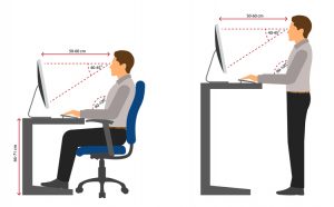
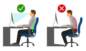
    Your monitor should be located directly behind your keyboard, and in front of you at approximately an arm’s length away. Adjust the top of the screen so it rests at or just below your eye level. Lower the monitor another 1-2 inches for ease of viewing if you wear bifocals. When reading images, try to reduce light as much as possible and focus on placing monitors on dark surfaces to help keep glare to a minimum.

  1. Chair position

    Be sure to select an office chair that supports your natural spinal curves and that feels comfortable to sit in. Adjust the height of your chair so your feet rest flat on the floor. If the height of your desk requires, use a footrest and place your feet flat on its surface. Adjust the armrests of your chair so you can comfortably rest your arms on the armrests while keeping your arms close to your body and your shoulders relaxed.

  1. Keyboard and mouse placement

    Place your keyboard and mouse on the same surface and make sure they are within easy reach. When using your keyboard or mouse, keep your upper arms close to your body, your hands at elbow height or slightly lower, and your wrists straight. Consider alternating hands to use your mouse or getting an adjustable mouse that responds to light touch for operation. Use keyboard shortcuts when possible to minimize how much you need to use your mouse.

  1. Key object placement

    Keep commonly used desk items within easy reach when you’re sitting. This includes items like pens, stapler, notepad, phone and other office items you use often when seated. If you’re not able to comfortably reach an item, it is better to stand up to retrieve an item rather than reach for it.

  1. Natural posture

    Many people have a tendency to start leaning forward when working from a desk or office chair. You want to ensure you are sitting back completely in the seat of your chair with your back against the back of the chair. This will help maintain the most natural posture for your body and back. For your legs, you want to sit so there is a 90° – 100° angle between your legs and back.

  1. Head and neck position

    The position of your head and neck is just as important as a position of the rest of your body when seated at your workstation. The head can tend to drift downward when working at a computer. This can cause pain in the neck, shoulders, and back. To avoid this, adjust the position of your monitor so your eyes focus on the middle of the screen. Add monitor arms to adjust monitors forward/backward and up/down when sitting or standing at your desk. Using a headset may also help keep the head and neck in a more relaxed and neutral position.

  1. Physical activity

    Having an ergonomic desk setup at home does not negate the need to get up and move—often. Experts recommend getting up from your desk and stretching, moving about your office, or grabbing something to drink every 25 to 28 minutes. If something doesn’t feel right or you feel uncomfortable, consider readjusting and reevaluating your desk setup. We offer free ergonomic consultations for home desks.

  1. Give your eyes a break

    In addition to moving your body, it’s also important to move your eyes off your computer screen approximately every 20 minutes, for 20 seconds, looking 20 feet away if possible, to give your eyes a rest (20/20/20 rule). Also, be sure your home office space is dimly lit and that there is no glare or reflection coming off your screen as you work.

Setting up an ergonomic workstation at home can take some time and effort – although the benefits such as fewer injuries, reduced strain, and increased productivity make it worthwhile.

To save radiology and medical professionals time and effort in setting up their own ergonomic home workstations, Double Black Imaging provides a custom workstation design service to meet your work needs at home. Our diagnostic imaging experts can make recommendations and help assemble every piece that’s needed for an ergonomic solution outside of the hospital or medical clinic.

Contact us for more information about our custom workstation design service.


References:

https://ehs.yale.edu/sites/default/files/files/ergonomics-home-office.pdf
https://www.mayoclinic.org/healthy-lifestyle/adult-health/in-depth/office-ergonomics/art-20046169
https://doubleblackimaging.com/product/anthro-steves-station/
https://www.ohsrep.org.au/offices_-_computers_and_eyesight

29 Apr 2020

Remote Radiology Monitors and Workstations

In this new COVID-19 era of working from home, attending Zoom meetings, and minimizing contact in the workplace, radiologists are finding themselves in a unique position; they now need to be able to read from almost anywhere.  Tools for at-home reading – including faster internet speeds, secure VPN connections, and Thin Picture Archiving and Communication Systems (PACS) clients – have made Teleradiology, or reading from home, commonplace. Flattening the curve and reducing your chances of contracting the virus are excellent reasons to consider adding or upgrading your home reading ergonomic workstation.

Where do we start? Regardless of location, we must seek out equipment that meets imaging requirements of the ACR, FDA, and DICOM standards, and that also significantly reduces strain on your eyes, neck, shoulders, and back. Consider the following as you examine diagnostic LED-backlit monitors, ergonomic configurations, workstations, or other equipment in the home office setting.

Requirements for Radiology Monitors

Radiology professionals risk missing key information, and also risk providing an incorrect diagnosis, if they are evaluating images and scans without the correct DICOM calibrated display. This is something that has been studied in depth for diagnostic imaging within the enterprise, and remains especially important for a remote office location. According to a white paper on Teleradiology published by the American College of Radiology (ACR), radiologists must use one high set of standards “for both Teleradiology providers and on-site radiologists”, as doing anything less could lead to compromised patient care. These standards cover display luminance, pixel pitch, and display size, and also ensure DICOM calibration.


Luminance

Luminance is a measure of how much photon, or light energy, reaches the eye, and is measured in candelas per meter squared (cd/m²). A spectrum of precisely calibrated luminance levels including brightest white, lowest black, and all levels in between, ensures subtle abnormalities are visible and discernible. According to ACR standards , the minimum calibrated luminance is 350 cd/m² for radiology modalities including X-Ray, CT, MR, PET, US, and NM, and 420 cd/m² for Mammography and Tomo. In addition to ensuring your remote reading display meets this minimum standard, it is also important to ensure the luminance output is stable over time. Medical displays have built-in backlight stabilization and monitoring. Our eyes don’t notice as a display slowly degrades over time or loses brightness. Backlight stabilization, a specification included in most medical grade displays, ensures the brightness level stays consistent over time, from monitor to monitor, and also controls the temperature at which the display runs.

Pixel pitch and display size

Individual pixels on a display determine the resolution of the monitor, with pixel pitch, or the density of pixels being a primary factor. Measured in millimeters from the center of one pixel to the center of an adjacent pixel, pixel pitch must be substantial in diagnostic monitors to provide the needed level of detail — approximately 200μm for most medical-grade displays.

Another way to think about resolution is to consider the matrix size, or pixels per millimeter of screen space. The recommendation from the ACR for diagnostic reading is 2.5 line pairs/mm minimum, equivalent to a 3 megapixel (3MP) 12.3” diagonal display. For mammography/tomography, 5MP dual-head displays or wide-format 8.8MP or 12MP single displays are the standards that typically carry FDA clearance for breast imaging.

Display size is also important to reduce the need for zooming in and panning out repeatedly. An appropriate diagonal display size is 21” diagonal for 5MP and 30-32” for large-format 8.8MP or 12MP displays.

DICOM calibration

Radiology displays used for at-home diagnostic imaging still need to meet the same standards they would inside Enterprise walls; they must be regularly calibrated to meet DICOM and ACR standards. Without meeting DICOM standards, diagnostic data is easily missed or misinterpreted – gray scaling and shadowing is truly lost.  DICOM calibration ensures the diagnostic data output from the display is perceptually linearized, enabling the human eye to discern all gray levels.  The user can be assured the diagnostic data is consistent on each display and from workstation to workstation throughout the enterprise.

Manual calibration with a handheld photometer and software combination is tedious and time consuming. Medical-grade displays used in the home office should ideally include built-in front sensors for luminance level calibration, rear backlight sensors to maintain stability continually and over time, and automatic calibration software. Calibration software should provide the ability to calibrate during convenient times (such as off-hours), send e-mail alerts for out-of-compliance issues, automatically diagnose and correct non-compliance issues if generate automated reports confirming adherence to the ACR, DICOM 3.14, and state specific requirements. A comprehensive, diagnostic quality hardware and software display combination is an invaluable tool for radiologists to feel confident in providing accurate diagnoses from a home office or remote location.

The DBI calibration software suite pairs your local display with a web based network administration option. Displays can be calibrated remotely and automatically to the DICOM grayscale display function and checked regularly for continued optimum performance.  Automatic e-mail alerting for nonconformance and report generation are also included.


Setting Up the Right at Home Radiology Workstation

In addition to ensuring displays meet ACR standards and produce the least amount of eyestrain, a radiologist should make sure his or her remote radiology workstation is set up with proper ergonomics in mind. Repetitive strain injuries are especially common with radiology professionals due to the hours spent in one position while viewing studies.

Countering the stress on the body from sitting in one place, engaging in repetitive motions to work a mouse and keyboard, or viewing monitors at an incorrect angle, is essential. The ergonomic workstation should include options for sitting or standing, the ability to adjust the monitor height or keyboard position, and also allow for multiple display configurations.

Lighting should be carefully considered due to the clear effects on diagnostic image representation. Excess ambient lighting reduces image contrast, creating a “washed out” appearance, thus reducing your ability to discern subtle luminance level differences on even the best monitors. Screen-glare contributes to eye fatigue and strain. Controlling ambient room lighting while adding proper task lighting are key to delivering the highest quality image performance with the most comfortable viewing experience.

Ergonomic Workstation Requirements


Confidence and comfort are necessary for Radiologists to successfully read from home. Valuable time adjusting workstations coupled with image quality concerns are counterintuitive to efficiently reading remotely or from home. Double Black Imaging can assist in designing the best home workstation to meet your individual needs. With cutting edge display technology, automated calibration software, and on-call customer service and support, we ensure your home office is quickly up and running.

Have a home workstation and find your workspace is not optimized ergonomically? We are happy to speak with you and create a custom solution that fits your equipment needs, space requirements, and budget.

Double Black Imaging provides ergonomic workstations and peripherals that ensure the right solution for your individual needs. With optimized ergonomics, your productivity will increase while eye strain and fatigue decrease.

Double Black Imaging has developed specially priced workstation bundles to assist radiologists with home office needs during this COVID-19 pandemic. Learn more here!


Contact the diagnostic imaging workstation experts today if you have questions about the best remote radiology monitor and workstation setup, including medical-grade displays, CPUs, and ergonomic desks.