Category: Medical Monitors

17 Aug 2022

Comparisons of LCD Color and Monochrome Displays in Diagnostic Imaging

The types and number of imaging data handled by hospitals are increasing as we witness improvements in medical technology.  This is having an impact on the type of imaging you need to use, as well as the type of display you need to view and analyze images.  As a result, you will see combined use of monochrome and color images to achieve the greatest diagnostic accuracy.

The choice between LCD Color and monochrome displays in diagnostic imaging is not a simple one.  Each has some advantages over the other, and a comparison that considers the unique needs of your facility is warranted.

For example, both color and monochrome displays can use grayscale to view images.  The grayscale that is used can impact the quality of the image and the accuracy of your diagnosis.

Which display you choose can determine how you use grayscale to view your images, whether you can switch from monochrome to color images, and whether images are converted manually by you, or through automated analysis.

Not only will these considerations influence the accuracy of your diagnoses, but the amount of time invested in achieving those outcomes.  Display functionality can save you time or cost you time, depending on your priorities and choices.

Comparing LCD color and monochrome displays for radiology is an important consideration when choosing the type of display to use in your hospital.


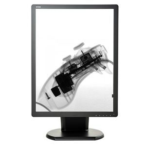
What Is An LCD Color Display


LCD stands for Liquid Crystal Display.  Today most monitors that radiologists use for reading studies are LCD.  These displays look like flat panel televisions and computer monitors; however, medical-grade displays meet a higher level of standards, have greater functionality for medical imaging, and are more robust for your needs.

Most LCD monitors use Light-emitting diodes or LEDs as the backlight source.  This is a change from past monitors, which used a Cathode Fluorescent Lamp (CCFL) technology.  LEDs are slimmer than CCFLs, and this allows the display to be slimmer than those with CCFL tubes.

Another important consideration is safety.  CCFL technology uses mercury which is dangerous to the patient, health care workers, and the environment.

LED technology is a mercury-free source of light.  While LED contains other dangerous metals, it doesn’t present the same health and environmental risk of mercury.  LED and LCD life expectancy is greater than the old CCFL.


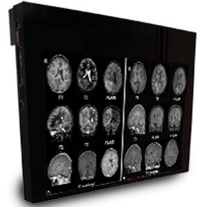
What Is A Monochrome Display?


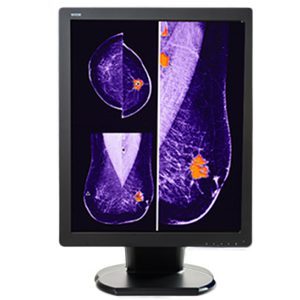
Traditionally, Monochrome medical-grade displays have been recommended in diagnostic radiology because they have a higher luminance.  The higher luminance of monochrome displays makes it easier to see the entire grayscale in an image.

More specifically, monochrome displays have only one color of phosphor.  Looking at the name, you can see that “Mono” means one and “chrome” means color.  Everything (text and graphics) is displayed in that color.

If you perform a lot of imaging modalities such as chest CT, DR, and other imaging modalities that require higher brightness and contrast performance, then monochrome displays are a great solution for you.

Monochrome displays are significantly more expensive than LCD color displays.  Less expensive monochrome displays are available.  However, they come at a cost of reduced luminance which impacts image quality.  Decreased image quality is a significant compromise when considering the impact on diagnostic accuracy.


How Are They Similar?


Both monitors require calibration to maintain accurate functioning.  Medical grade displays are usually equipped with controls to facilitate grayscale calibration.  Monitors need to maintain calibration to the DICOM standard.  Maintaining calibration is particularly important when images need to be compared to previous images of the same person and condition on a different display.

Both LCD Color and Monochrome displays should be medical grade because they are designed for high volume use and facilitate viewing radiological images with the detail needed for accurate diagnosis and efficient completion of the analysis.

There can be some variability in uses that are recommended; however, based on the general characteristics of LCD Color and Monochrome displays are recommended for several of the same modalities.

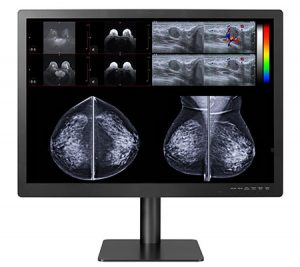
For example, looking at LCD color and monochrome displays used for mammography and tomography, both are recommended for Computed Radiology (CR), Computed Tomography (CT), Digital Radiography (DR), Ultrasound, PACS and Mammography.

They come in similar sizes with similar numbers of pixels and resolution.  The viewing angle for these displays is not significantly different.  There is no difference in palette, video inputs, dimensions, and weight, and both have external power supplies.


How Are They Different?


Monochrome displays have a much higher cost. The trade-off is the longer life span of the display.  Also, they have higher luminance which facilitates efficiency and accuracy.

Color monitors have a lower maximum luminance compared to monochrome displays.  This doesn’t necessarily mean that diagnostic accuracy will be decreased.  The study by Geijer, et al.1, indicated that accuracy with color LCD and Monochrome monitors was not significantly different, but that the dwell time for color monitors was greater.

All displays can be windowed.  However, color displays often need to be windowed to improve image quality for specific area you are analyzing.  While a normal part of reading a digital image, it is more necessary in viewing on color displays, and this can reduce efficiency and limit the ability to compare objects in the window with those restricted from view.

Color and monochrome images should be viewed using different grayscales.  Monochrome images are best viewed using the DICOM 14 Grayscale Standard Display Function (GSDF) while color images are best viewed using Gamma 2.2.

In addition to modalities recommended for both displays above, LCD color displays are recommended for Nuclear Medicine, 3D, Positron Emission Tomography (PET), and Tomosynthesis.  LCD Color displays consume more power than monochrome.

An advantage of color displays is that they can show color information.  As medical technology advances and more modalities use color, this is an important consideration.  Modalities that already use color include Doppler ultrasound, 3D reconstructions in computed tomography (CT), functional magnetic resonance (MR) imaging, and nuclear medicine, including PET.

Given that color displays are significantly less expensive, they can be replaced up to four times more often within a fixed budget1.

Meanwhile, monochrome displays have a higher contrast ratio and brightness than LCD Color displays.


Selecting The Right Display For Your Office


Displays now function to read images and adjust between color and monochrome, or to allow easy switching from one functionality to another.  This makes the less expensive, LCD Color displays more attractive.  However, there are circumstances when the monochrome display is the better choice.

Which display you choose will be influenced not only by cost, but also by the current and future needs of your hospital, which is determined by the types and numbers of modalities you perform as well as what you anticipate you might add to the modalities you perform during the life of the display.


Get Your Radiology Displays From Double Black Imaging


Double Black Imaging understands the importance of displaying high quality images while containing costs.  We can help you choose the best display options for your facility and your budget.  We offer a range of options that are the best LCD Color and Monochrome displays on the market.  Contact us today to set up a discussion about how we can help you with your next display purchase.


1 Geijer H, Geijer M, Forsberg L, Kheddache S, Sund P. Comparison of color LCD and medical-grade monochrome LCD displays in diagnostic radiology. J Digit Imaging. 2007 Jun;20(2):114-21. doi: 10.1007/s10278-007-9028-5. PMID: 17340227; PMCID: PMC3043910.

12 May 2022

What Are Surgical Displays And Monitors?


Surgery monitors and ER monitors provide high quality images that facilitate your ability to successfully perform surgeries and procedures. A quality monitor allows you to see the patient’s anatomy in detail and make diagnostic and treatment decisions for the best patient outcomes.

Medical-grade monitors are used in operating rooms, emergency rooms and medical offices.  Displays you use in image-guided procedures such as endoscopy (gastrointestinal, arthroscopic, laparoscopic, or robotic guided surgery) and interventional radiology are usually referred to as surgical displays or surgical monitors.  You can use them to display a variety of critical information, including patient data, x-rays, vital signs, brain activity, endoscopy images, PACS images, surgical feeds, etc.


Types of Surgical Displays


Surgical displays come in a variety of forms, shapes, and sizes.  These devices include displays you can use near a patient (near-patient displays), large screen operating room displays, user interfaces (touch displays), and control displays which are often used in interventional radiology.

Surgical displays can offer high-definition or 4K visualization for presented visual images.  These displays can be mounted on your radiology practices wall, on an equipment column, or on a surgical light arm.


Diagnostic Displays


As a physician or radiologist, use a diagnostic display to view various diagnostic images like Computerized Tomography (CT) scans, Magnetic Resonance Imaging (MRI), and X-rays.

Medical facilities are relying on diagnostic displays more as new procedures require you to employ endoscopic visualization to perform surgery or diagnostic procedures.

High Definition and Ultra High-Definition Surgical Displays


As you rely more on image-guided surgical devices, you can appreciate the movement from High Definition to Ultra High Definition (4K) technology incorporated into new systems and monitors.  A 4K monitor provides four times the image detail compared to High-Definition images. This means that you can see more anatomical detail and more clinically relevant information in order to provide the best service to your patient.

Another benefit of 4K displays is that they provide a wider color gamut.  It provides you with a broader spectrum of reds – which is especially helpful in the OR.  It also provides more detail and color-corrects images, which means you can differentiate tissue types and structures such as nerves, blood vessels, organs, and organic material within your patient.

It is also important to note that with the detail gained with 4K resolution, you gain a better holistic perception and a more immersive environment to operate with greater precision.


Large Format Displays


Simply stated, Large Format Operating Room displays give you a larger viewing area.  They can be mounted on the OR wall so that your entire team can view the surgical site during the procedure.

This feature makes large format displays ideal for use in your hybrid ORs, interventional X-ray and cardiovascular suites.


Important factors in a Surgical Display


           Crisp Image

Clear, crisp images mean you won’t have artifacts and visible pixels.  So, you can see the fine detail of the surgical site.  Some surgical displays offer multi-image visualization for viewing multiple images simultaneously in high quality.

The backlight stabilization of surgical displays ensures consistent image brightness and maintained focus for moving images.

Finally, you can see details clearly, even when you are not standing directly in front of the display.  So, there is no need for frequent readjustments of display location and angle.

          Fan-less Design

A fan-less monitor has the advantage of being easier to clean and disinfect, preventing the spread of contaminants and saving your staff time and resources.

Also, the fan noise is eliminated, enabling your team to communicate more effectively, and reducing concerns about errors related to noise interference.

          Ventless Design

Surgical monitors with vents require additional time and effort to clean. You risk contamination of your hands, the patient, or surfaces near the patient if the pathogens are not completely removed by thoroughly cleaning all surfaces.

A ventless design has two great advantages:

  1. It reduces the surface area that you must clean, making cleaning easier and less time consuming.
  2. It also reduces areas where dust can settle, and bacteria can grow.


          Impact-resistant Design

Surgical monitors with impact-resistant shields protect the panel from physical damage, which means your technology investment is protected against collisions with lights, IV poles, and other structures in the OR.

This is particularly important when your staff is working quickly to accommodate frequent turnovers for new procedures.

         Medical Grade Power Supply

A Medical grade power supply has a long lifecycle and minimizes the risk of shock.  Also, you gain protection against a technical or power failure so you can maintain critical information during a procedure.


Why are Double Black Imaging Surgical Displays the Best?


Double Black Imaging is committed to providing you medical imaging technology that enhances your diagnostic capabilities.

Here are just a few of the many reasons Double Black Imaging Displays are the best:

  1. Our displays provide bold, crisp images even when you view them from a distance.
  2. We provide you with the world’s most technologically advanced Surgical Displays.
  3. They are built to withstand years of use.
  4. Our monitors give you unmatched quality, clarity, and consistency.
  5. We offer a full line of splash-proof Surgical Operating Room Displays.
  6. Our solutions meet and exceed all surgical imaging requirements.
  7. Our displays offer full HD resolution, ultra-high brightness, and
  8. They support a wide variety of inputs so you can interface with your new or technologies and eliminate costly signal conversion, signal degradation and image lag.
  9. Our monitors to deliver exceptionally fast response time when viewing live video or motion, with in versatility for picture in a picture or picture by picture format.
  10. We guarantee that every display has certification for classification as a medical display.


Why Choose Double Black Imaging?


At Double Black Imaging, we base each product and business decision on how to best serve your needs.  We strive to build a long-term relationship with you by offering the most advanced equipment at competitive prices.

Double Black Imaging began in 2002 with the goal of making medical imaging more efficient and improving healthcare.  We maintain that standard by providing the best value in the industry and by offering technologically advanced medical imaging solutions that enhance diagnostic capabilities, along with exceptional customer service and support.


Premier Customer Service


Double Black Imaging’s commitment to making the best decision for you, our customer goes well beyond the sale of OR Monitors.

Surgical Displays and Monitors


Our industry-leading customer service means we provide you the necessary knowledge and support, before and after your purchase.

To learn more about your ideal OR Monitors or our other products and services, contact us today at (877) 852-2870 or email us at sales@doubleblackimaging.com.

20 Apr 2021

Prioritizing MRI Safety with Safe Imaging

The evolution of MRI safety has become more challenging than ever for radiologists, MR technologists, and referring physicians.

As Sammet explains” The increasing clinical demand for Magnetic Resonance Imaging (MRI) with its superior soft-tissue contrast compared to other radiological imaging modalities and potential physiological and functional applications has contributed to the installation of almost 30,000 MRI scanners worldwide.”

Combined with this is the rapid increase in the number of patients who have implants due to advancements in medical implant technology.

This growth in clinical demand and the number of MRI scanners installed has required that significantly more health professionals be educated about MR safety to protect patients, themselves, and others who may be exposed to the MR environment.

What Is MRI and What is it Used For?

Magnetic Resonance Imaging (MRI) is a non-invasive imaging technology that produces detailed three-dimensional images of the body’s anatomy. The technology works by stimulating protons in the water of living tissues and then identifying how the protons’ rotational axis changes.

This is done by using extremely strong magnets that create a strong magnetic field forcing protons in the body to align with the magnetic field. By pulsing a radiofrequency current through the body, the protons are forced to spin out of balance as they strain against the pull of the magnetic field.

Then the radiofrequency is turned off allowing the protons to realign with the magnetic field which releases energy that the MRI sensors detect. How long it takes for the protons to realign with the magnetic field and the amount of energy released when this occurs differs depending on the chemical composition of the molecules and the environment. Using these measurements, physicians can identify the different types of body tissue.

Physicians use MRI to identify differences between healthy and unhealthy tissue in the body. It has become the preferred imaging procedure for diagnosing potential medical problems. Many body tissues can be assessed using MRI including the heart, blood vessels, internal organs, abdomen, pelvic area, brain, spine, breasts, and joints in the body.

How Safe is MR Technology?

Millions of MRIs are performed in the US each year. And according to the FDA, approximately 300 adverse events are reported from healthcare facilities, distributors, manufacturers, and patients related to these scans. Overall, this demonstrates the safety of MR technology for patients and healthcare personnel when the appropriate safety precautions are implemented and correct equipment is used.

Why Has MR Safety Become More Challenging and Complex?

MR technology has become increasingly complex not only because more scanners are being used and more procedures are being done, but also because scanners with higher field strengths are being developed and used in hospitals, institutions, and private practice.

In addition, radiologists are not required to have any formal training about MR imaging physics or MR safety concerns related to this form of imaging as Emanuel Kanal, MD, director of magnetic resonance services at the University of Pittsburgh explains.

The complexity and challenges related to MR safety are due primarily to three factors:


  1. The sophisticated technology that makes MR imaging possible


    The static magnetic field of an MRI machine is incredibly powerful. For example, a 1.5 T magnet exerts a magnetic force that is 21,000 times greater than the earth’s magnetic field. With this force, small metal objects such as hairpins and paper clips can be made airborne and move at speeds of up 40 mph into the machine, injuring a patient and anyone in the path of the flying object.

    Larger medical objects made of stainless steel such as scissors, hemostats, clamps, as well as oxygen tanks, metal gurneys and even floor buffing machines can become projectiles and injure people in the room as well.

    For this reason, all metal objects regardless of their size must be kept out and away from the room housing the MRI machine.

    Biological risks including cell growth and aberration and some reversible effects such as headaches, irritability, fatigue, and hypotension have been noted in human subjects exposed to static magnetic fields of 2.0 Tesla and above. Most MRI equipment currently produces fields less than 2.0 T which have not produced any known biological effects. However, the FDA recently approved the first 7 T MRI system for use in the US in October 2017 so these risks may now exist.

    One of the risks related to the radiofrequency (RF) field is the risk of current-induced thermal burns to the patient’s skin. This can occur if the RF field produces currents in wires that are near or attached to the patient. The RF field may also produce currents in intra-cardiac leads causing unintended cardiac pacing. Lengthy imaging procedures can also increase core body temperature although this is primarily a concern in infants.

    Cryogenic risk exists if the magnetic field is shut down accidentally or for planned maintenance. When this occurs, liquid helium inside the magnet begins to turn into a gas that may seep into the scanning room and displace oxygen leading to possible asphyxia of the occupants.


  2. Patient variably


    One of the greatest concerns about scanning patients is if they have implants, what material these implants are made of, and how these implants may react when exposed to the MR field. While the FDA allows manufacturers to market certain implants as “safe” for MR imaging “at or up to certain conditions under which they were tested”, MR environments are highly variable and may exceed these test parameters.

    According to Kanal, this variability may result due to different strengths of the MR energies at different positions in the MR scanner and scan room as well as at different positions throughout the patient’s body. And “How they vary can and do differ with different MR scanners and gradient coils and RF transmitting coils among varied scanners of the same or different MR manufacturers.”

    For these reasons, Kanal developed a smartphone app called MagnetVision that has all this information built in so it is able to calculate in real-time the effect of MRI on a particular implant, coil, MR scanner or patient.


  3. The increase in imaging procedures performed

    The rapid increase in MRI technology and software has also led to new imaging procedures being developed including multi-contrast imaging and lung imaging. With new procedures and technology come issues of safety that must be evaluated, documented, and communicated to those using this new technology with patients.

    When these potential MR safety issues are understood, proper safety protocols are followed, and the correct equipment is used, MRIs are safe and extremely effective in producing exceptional images that contribute to timely patient diagnosis and treatment.

    To meet our customers MR safety needs, Double Black Imaging offers two different sizes of MR Safe Displays that are fMRI compatible and provide a 2.3-megapixel color LED backlit display. They are available as 24-inch and 32-inch LCD displays to accommodate varying space constraints and bore sizes.

    Both LCD medical displays provide lag free output and no interference with the scanner even when positioned at the bore exit. Display specifications include an extraordinary spatial spatial uniformity of 2% over the central 75%, ultra-high brightness of 1000 cd/m2, and a contrast ratio of 1000:1 resulting in unparalleled MR image quality.

    At Double Black Imaging, we understand the challenges technicians and radiologists face in producing high quality images while keeping patient and healthcare personnel safety as their top priority. Contact us to learn how our MR Safe displays can help meet your practice’s imaging needs.


Source List:

https://www.ncbi.nlm.nih.gov/pmc/articles/PMC4848040/
https://www.itnonline.com/article/recent-advances-mri-technology
https://www.nibib.nih.gov/science-education/science-topics/magnetic-resonance-imaging-mri
https://www.ismrm.org/resources/information-for-patients/
https://www.mayoclinic.org/tests-procedures/mri/about/pac-20384768
https://www.fda.gov/radiation-emitting-products/mri-magnetic-resonance-imaging/benefits-and-risks
https://www.diagnosticimaging.com/view/qa-tackling-complexity-mr-safety
https://physicsworld.com/a/mri-safety-an-urgent-issue-for-an-increasing-crowd/
https://radiology.ucsf.edu/patient-care/patient-safety/mri/potential-hazards-risks
https://www.itnonline.com/content/fda-clears-first-7t-mri-system-magnetom-terra
https://www.itnonline.com/article/recent-advances-mri-technology

23 Mar 2021

WHY TRANSITION TO RADIOLOGY READING FROM HOME?

Radiology from Home

Since the widespread emergence of COVID-19 in North America in early 2020, remote radiology reading from home (also known as teleradiology) has seen a surge in popularity. Teleradiology refers to radiologists receiving images to interpret from a facility in a different geographical location than where the radiologist performs the reading. This may be in a country in a different time zone halfway around the world or in the radiologist’s home located just blocks from the hospital they are contracted with.

What’s the incentive to shift to remote radiology reading?

Social distancing requirements to help stop the spread of the virus during the pandemic has required a radical shift in how radiology is practiced in hospitals and healthcare centers.

Mohammed Imran Quraishi, MD, assistant professor of radiology at the University of Tennessee, and colleagues surveyed 290 geographically diverse institutions to determine how many sites were transitioning to teleradiology as a result of the pandemic. They received 174 responses and found a significant number of sites had radiologists installing workstations at home (65.2%) and a sizable movement to internal teleradiology (73.6%) to replace daytime shifts.

Beyond the safety related reasons for shifting to more remote radiology reading, performing radiology from home offers increasingly strong personal and economic incentives for physicians as well.

Jackson notes “When teleradiology first entered the industry as a career option, it was pigeon-holed into being an overnight service only. Radiology practices used teleradiology as a way to avoid overnight call. However, in recent years, reading images remotely has ballooned as a viable career option for radiologists who simply don’t want to work in hospitals or imaging centers.”

And if the increased flexibility of performing radiology from home is not enough to convince radiologists to consider working more at home both during and after the pandemic, the economic trends in the sector might be.

In February 2020, Grand View Research, an India & U.S. based market research and consulting company, reported the size of the global teleradiology market is expected to hit $10.9 billion USD by 2027. Growth in this sector is expected to be driven by a shortage of radiologists paired with an increased demand for imaging procedures.

The fastest growth is expected to occur in CT. This is due to quicker testing, widespread use, high-resolution image quality and the arrival of newer 4D generation systems.

In 2019, x-ray had the largest share of the teleradiology market due to its broad scope of application from identifying bone fractures to dental injuries. Increased demand for core diagnostics and imaging at the bedside combined with new technologies is predicted to drive growth even more.

    1. Flexible work
      Michael Yuz, MD, an executive radiologist with USARAD, a radiology-on-demand company points out, “As the opportunities have expanded, so have the positives that come with the work.” However, he is quick to point out that while radiologists working from home can choose their days and hours of work, they will not end up working less. In fact, he says it’s not unusual for radiologists working from home to work twice as many hours as their colleagues practicing in hospitals or imaging centers and to routinely put in 12-14 hour workdays.


    1. Better work-life balance
      Being able to choose what days and hours one works and the ability to work at home allows teleradiologists to have more freedom to decide where they want to live. It also allows them to spend more time with family and friends. This results in radiologists who are happier with where they work and live and as a result, they experience less work-related stress according to Michael Rothman, MD, a teleradiologist based in Bethlehem, PA. Quraishi et al. confirmed this in their study stating 64.8% of respondents who transitioned to working from home during the pandemic reported decreased stress levels. And this occurred while 96% of respondents found an improvement or no change in turnaround times for readings. Michael P. Recht, MD, Professor and Chairman of NYU Langone Department of Radiology “said he is ‘very much’ in favor of remote reading and noted that about one-quarter of his faculty took part in this practice—thanks to a wellness initiative—prior to COVID-19.” He says almost 70% of his radiologist staff have taken part in the program during the pandemic with these physicians experiencing improved work-life balance due to the increased autonomy and flexibility in their work.


    1. Ability to select a subspecialty
      As Yuz explains, most imaging facilities expect their radiologists to function as generalists. However, teleradiologists working from home have the ability to choose and focus on a subspecialty. They may still be required to perform some generalist work but they can also work within their chosen specialty as well. And depending on where they live, teleradiologists may be able to go into local hospitals and perform readings for hospital radiologists who lack experience in a particular subspecialty.


    1. Ability to select partners
      Physicians who transition to remote radiology reading also have the opportunity to choose what hospitals and imaging centers they partner and work with. This means being able to work with smaller or larger centers (depending on preference) as well as radiology centers offering imaging to patients in specific subspecialties the radiologist may want to focus in.


    1. Reduced dependence on external/contracted readings
      In the research by Quraishi et al., they also found that as more radiologists transitioned to working from home during the pandemic, many radiology practices saw a corresponding drop in the need for external or contracted readings. This finding was common across the U.S. as a result of fewer non-essential cases and lower case volumes overall.


What will you need to make remote radiology reading successful?

While performing radiology from home has its benefits and can be a very rewarding way to work in the profession, it can also be extremely frustrating and next to impossible without the right equipment and support.

    1. PACS equipment

      Not surprisingly, having up-to-date technology that is suitable for imaging and patient information security according to the ACR–AAPM–SIIM Technical Standard For Electronic Practice of Medical Imaging guidelines is a must. Most home workstations will require at minimum multiple diagnostic quality monitors, a microphone or dictaphone for speech recognition, and a computer with PACS access that is secure. An ergonomically designed workstation is an important feature to consider as well to help reduce fatigue and repetitive strain injuries.  Double Black Imaging has released teleradiology bundles that can be easily customized with CPUs and ergonomic workstations including seating.  Our configuration team ensures that each workstation is preconfigured and labeled for ease of use at the end user site.


    1. Fast internet connection

      Sammer et al. found an internet connection of 80 Mbps over the hospital VPN provided radiologists working at home on PACS workstations with nearly the same results as working in-house but noted that speeds decreased during peak work hours when more people were working from home. Speed was also reduced if the internet connection was being used by someone else concurrently at home. They reported internet speeds as low as 30-40 Mbps were “acceptable” but this resulted in noticeable lag while speeds of 200+ Mbps allowed for a “seamless experience”.


    1. HIPPA compliance

      To practice teleradiology, radiologists working from home must ensure they are working on a secure network to protect patient information and privacy. One important way to protect patient information when working remotely is via two-factor authentication when signing onto PACS at home. While it may take an extra couple seconds to do this, it’s essential to ensure patient data is not compromised.


    1. Integration of and access to patient information

      Radiologists working from home must have access to patients’ electronic medical records (EMRs). This includes access to previous imaging exams and reports.


    1. IT support and self-help instructions

      Having the right workstation setup and internet is essential. But it’s just the start of what’s required for radiology reading from home. Technology is great—when it works. When it doesn’t, radiologists working from home need access to 24/7/365 IT support when problems arise. As Matthew Hayes, a PACS manager at Radiology Partners explains “Aside from the IT infrastructure challenges required for at-home PACS, organizations need to consider who is going to troubleshoot problems. What if a head CT for a stroke protocol is taking 15 minutes to load? The radiologist needs to talk with someone quickly via a helpdesk. Even simple fixes such as HDMI connections can be solved ahead of time with simple, easy-to-read PDF instructions” he said.


    1. Licensing and legal support

      For physicians performing radiology from home, Yuz and Rothman explain radiologists may be working with many different hospitals and imaging practices across the country and even internationally. This will require licenses in all the states and countries a physician works with as well as legal representation to ensure liability coverage.


The diagnostic imaging experts at Double Black Imaging can help you select a home workstation that suits your needs and budget during COVID-19 and beyond. Contact our radiology display experts today with your questions. We’re eager to help any way we can.

Contact our diagnostic imaging experts


Source List:

https://collaborativeimaging.com/the-pros-and-cons-of-teleradiology/
https://www.healthimaging.com/topics/imaging-informatics/teleradiology-adoption-spiked-covid-19
https://www.ncbi.nlm.nih.gov/pmc/articles/PMC7245278/
https://www.diagnosticimaging.com/view/teleradiologist-experience
https://www.grandviewresearch.com/press-release/global-teleradiology-market
https://www.healthimaging.com/topics/imaging-informatics/remote-reading-radiologys-virtual-future
https://www.acr.org/-/media/ACR/Files/Practice-Parameters/Elec-Practice-MedImag.pdf
https://www.arrs.org/downloads/ajr/COVID-19-WFH.pdf
https://www.healthimaging.com/topics/conferences/remote-reading-here-stay-how-radiology-can-prepare-virtual-future

24 Feb 2021

Section 179 Tax Deduction

Section 179 Tax DeductionTax time will be here before you know it. Now is a good time to start pulling together your business expenses and documentation from this past year to ensure you are capitalizing on all the tax deductions you are entitled to as a business owner. The Section 179 tax deduction is an important tax saver for business owners that should not be overlooked.

This deduction can and does change from year to year (and has even changed within a tax year). Therefore, it is important to be aware of your deduction limit each year including the bonus depreciation which may also change depending on the tax year.

What is the Section 179 Tax Deduction?

The Section 179 tax deduction is a section of the IRS tax code originally designed to provide incentives and tax relief for small businesses although large businesses may also benefit from this deduction.

Section 179 “allows businesses to deduct the full purchase price of qualifying equipment and/or software purchased or financed during the tax year. That means that if you buy (or lease) a piece of qualifying equipment, you can deduct the FULL PURCHASE PRICE from your gross income.” This U.S. government incentive is designed to encourage businesses to buy the equipment they need while investing in themselves and their businesses.

Who Qualifies for the Section 179 Tax Deduction?

For the 2020 tax year, the good news is all businesses that finance, lease, or purchase used or new equipment outright totaling less than $3,630,000 should qualify for the Section 179 deduction.

More specifically, $1,040,000 of business assets qualify for the complete Section 179 deduction in 2020. That amount is reduced dollar for dollar when the amount of qualified assets used for business purposes reaches $2,590,000.

What Requirements Must Be Met to Claim the Section 179 Deduction?

Any equipment, vehicles, or software claimed as a tax deduction under Section 179 that is used for both personal and business purposes (such as cell phones for example) must meet the “More Than 50 Percent Business-Use” requirement. This means that the equipment, vehicles, or software being claimed must be used for business purposes more than 50% of the time that it is used for it to qualify for the Section 179 tax deduction.

What Equipment Qualifies for the Section 179 Deduction?

The following equipment “generally” qualifies for the Section 179 deduction and may be new or used—however it must be “new” to you as a business owner for it to be claimed as a deduction. It must also be purchased (and used) between January 1 and December 31 of the tax year in which you are wanting to claim it as a tax deduction against your business. The term “purchased” includes equipment you have leased, financed, or purchased outright with your own funds.

Equipment that generally qualifies includes:

  • •Machines and a variety of equipment purchased for business use such as computers and “off-the-shelf” computer software.
  • •Equipment purchased and used for business and personal use (the section 179 tax deduction is based on the percentage of time you use this equipment for business purposes).
  • •Office equipment and furniture.
  • •Different types of tangible personal property used for business purposes.
  • •Specific types of improvements to existing non-residential buildings such as roofing improvements, fire suppression installations, security and alarm systems, and heating, ventilation, and air conditioning improvements.
  • •Non-structural equipment and property attached to the building where you do business.
  • •Vehicles used for business purposes (gross vehicle weight must exceed 6,000 lbs).


What’s the Difference Between Section 179 and Bonus Depreciation?

Bonus depreciation is taken after the Section 179 deduction is applied. Bonus depreciation usually benefits larger businesses that exceed the spending cap for new capital equipment in a given tax year (currently $2,590,000 for 2020).

It is also important to note that bonus depreciation is not offered every tax year. However, it is available for the 2020 tax year at a rate of 100%.

The most notable change for bonus depreciation in 2020 is that it may be applied to both new and used equipment, as long as the equipment is new to the business owner. This is a change from previous years in which bonus depreciation could only be applied to new equipment.

Businesses that experience a net loss this past year still qualify to deduct some of the cost of the equipment they purchased and then they may carry-forward the remaining loss to the following tax year. Similarly, if a business has no taxable profit during this current tax year, it may carry forward the loss experienced on equipment purchases to the subsequent tax year.

Is There a Way to Maximize My Tax Savings with Section 179?

Yes there is! One of the best ways to maximize your tax savings in 2020 is to lease or finance equipment purchases according to the Section 179 Qualified Financing rules. This is because as a business owner, you will only have to make the required lease or financing payments for the equipment during the year. However, you are entitled to deduct the full purchase price of the equipment you purchased in 2020, regardless of whether it is new or used.

In effect, financing or leasing equipment and then taking the full Section 179 tax deduction can easily help business owners save more money in taxes than they spent in finance or lease payments throughout the year.  Double Black Imaging offers a variety of leasing and financing solutions for our display systems, ergonomic workstations and CPU options!

Is It Possible to Preserve the Cash Flow in My Business and Still Take the Section 179 Deduction?

Absolutely! This has been and continues to be a year filled with uncertainty. Protecting cash flow for unanticipated expenses is a wise business idea. As discussed above, choosing to purchase equipment by leasing it can also put more tax dollars back in your business.

To keep lease payments as low as possible to preserve cash flow, consider asking about a non-tax capital lease. This type of lease will allow you to make smaller payments, protect your cash flow, and allow you to write-off the full price of the equipment you purchased up to the deduction limit— all while making the smallest payments possible.

How Do I Calculate My Potential Savings Using Section 179 on My Next Purchase?

That’s a great question. Fortunately, Section 179.org has an easy calculator that can help you estimate your potential savings if you are considering buying equipment this year.

To give you an idea of just how significant the tax and equipment savings can be for a business owner, Section 179.org provides this example:

“Using a $75,000 equipment cost for a sample calculation shows how taking advantage of the Section 179 Deduction can significantly lower the true cost of the equipment purchased, financed or leased. In our example, $75,000 in equipment purchased has a true cost of $48,750. That’s $26,250 saved. Would you like an extra “25 grand-plus” this year on equipment you needed anyway?”

How Do I Ensure I Get the Section 179 Tax Deduction?

It is important to know you must elect to take the Section 179 tax deduction for business equipment you have purchased, leased, or financed during the calendar year. It is NOT automatically applied to your tax return.

To elect to take the Section 179 deduction, complete Part 1 of Form 4562 from the IRS and attach it to your tax return. Or speak with your tax preparer and tell them you want to elect to take the Section 179 deduction.

In addition, the Section 179 deduction is taken on an “item-by-item” basis. Therefore, you can choose to claim the deduction for only some of the equipment and/or software you purchased and not all of it if you choose.

Can You Use Section 179 Every Tax Year?

Yes, the Section 179 tax deduction can be used for business expenses and depreciation every year that you incur these expenses in your business. However, it is important to keep in mind that changes are made to Section 179 every year, and sometimes throughout the year. Therefore, be sure to stay informed about these tax code changes, what equipment, property, and improvements qualify, and what deductions you might be entitled to.

Be sure to speak to your tax preparer if you are not sure what deductions you are entitled to or prior to making large equipment or software purchases for your business to determine the most tax efficient way to do this.

Double Black Imaging provides business owners with medical displays for PACS, monitors, and software as well as ergonomic workstations that qualify for the Section 179 deduction. Contact us at (844) 879-2247 with your business needs today and be sure to tell our diagnostic imaging specialists you want your purchase to qualify for the Section 179 deduction.

Contact our diagnostic imaging experts


Source List:

https://www.section179.org
https://www.irs.gov/pub/irs-dft/i4562–dft.pdf
https://www.crestcapital.com/section_179_bonus?_ga=2.120781388.519583165.1608581164-847429404.1608581164