What is DICOM in Medical Imaging?
DICOM stands for Digital Imaging and Communications in Medicine. It is an internationally standardized protocol for managing and transmitting images with related data. It ensures interoperability of systems that produce, display, query, retrieve, share, send, store, print, and process medical images.
Why Is DICOM in Radiology Important?
Advances in imaging technology and increased use of computing in clinical work created a need for a standard method of transferring images and their associated information. DICOM was developed to answer this need. It can ensure this transfer between devices, regardless of vendor or manufacturer.
DICOM in radiology is important to maintain this accuracy because facilities rely on medical imaging for accurate communication and profit.
With DICOM it is easier for physicians to access images and reports, allowing access from in house or remote locations. This greatly improves the efficiency, and therefore the quality, of patient care.
What Does A DICOM File Contain?
DICOM images consist of two components: a header and the image.
The header contains the data that describes the image, primarily patient data. As with other medical files, this includes the patient’s demographic information such as their name, age, gender, and date of birth.
The header can also contain information about the image, such as acquisition parameters, pixel intensity, matrix size and dimensions of the image.
How To View A DICOM File
It is only possible to view DICOM images using special software mean specifically designed for viewing these files. The software can be proprietary or third party.
Proprietary software is what is installed in the equipment you purchase for medical imaging.
You can view the images at the same workstation where the images, such as CT or MRI, were acquired by the machine using the installed DICOM viewer. It will allow you to view images sequentially and to reconstruct the images.
Unfortunately, the proprietary software only allows you to view the images in the same location as the hardware. The only way you can transfer the images is to a portable storage device or a network; however, you need to compress the images to do that. Compression and exporting images usually prohibits access to the original image.
Because of these and other issues, third-party software has been developed and is used more commonly. Third-party applications allow you to open DICOM files from any source (PACS server, the internet, a CD, or a DVD).
There are many viewing applications available for DICOM in radiology. Each viewer has different features, and you can consider the features available in each application as they compare to your viewing needs. The availability of third-party applications facilitates interoperability of your medical viewing device with other devices.
What Can I Do To A DICOM File In Addition To Simply Viewing The Image?
DICOM medical image viewers provide functionality that can increase the efficiency and effectiveness of your work. Some of the capabilities go beyond what was first imagined when imaging technology was developed. Among the things you can achieve with DICOM are:
- Comparison of medical images – With DICOM, you can view two digital images side by side to make comparisons.
- Enhancement of image quality – DICOM allows you to zoom in to see detail and to change the contrast in an image. By increasing or decreasing the brightness of the image, you can make better distinctions between radiodensity and radiolucency in different areas of the image.
- Reconstruction of images – This allows you to see aspects of the anatomy that are not available from the original images.
- Two-dimensional to three-dimensional rendering is a method that takes the initial two-dimensional images from the initial DICOM data set that are taken in all three planes (axial, coronal, and sagittal) and reconstructs them into a three-dimensional rendering.
- Multiplanar Reconstruction (MPR) uses the 3D reconstructed images and makes slices of them to allow you to view different anatomical levels at different angles than those acquired in the initial images.
- Combination of images – Some DICOM applications allow you to combine different modalities, such as PET and CT images. Using this combined image, you can simultaneously leverage the advantages of both types of imaging modalities. So, for example, a PET scan would locate areas of high metabolic activity, and you could combine that with the CT scan to map it to corresponding anatomical sites.
- Measurement of image targets– If you want to measure the linear size or volume of anatomic structures, you will appreciate apps that have this functionality. Being able to make these measurements accurately and efficiently is useful in assessing treatment efficacy or in planning treatment.
Can You Transfer A DICOM File Into Other Formats?
Each DICOM file contains several high-resolution images. Because this can create a very large file, compression is needed so you can transfer it.
It is important to note that compression of DICOM files uses two strategies: lossless and lossy.
Lossless does not lose any information when it is compressed. You can easily recover the original file at any time. However, this requires a lot of processing and results slow file opening and saving. A lot of compression is not possible with this strategy.
Lossy compression achieves greater compression because it removes some data. Typically, redundant data is removed. However, if a significant amount of compression is done, image quality can be impacted.
DICOM files can be compressed and exported into various formats, including JPEG, TIFF, PNG and GIF. This table summarizes the formats.
Other Capabilities of DICOM in Radiology
In addition to capabilities that enhance your viewing of and working with images, DICOM also has several functionalities that enhance the operation of your hospital or clinic.
DICOM supports network image management, network image interpretation management, network print management, image procedure management and offline storage media management.
DICOM simplifies the way you interface with your imaging equipment and can often be achieved by a “plug and play” system.
The system also works to group images with similar properties, which facilitates storage and retrieval.
Workload is also minimized by communication between Imaging Equipment and Existing Information Systems. Once connected, DICOM generates a list of scheduled imaging procedures for that equipment. Therefore, DICOM eliminates the need for duplicate data entry at the imaging equipment console. Furthering efficiency, DICOM in radiology allows the user of that equipment to query another system and obtain additional details about the procedure and/or the patient.
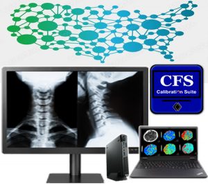
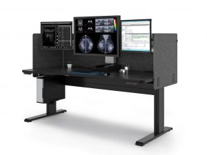
Double Black Imaging Is Here For All Your Medical Imaging Needs
Double Black Imaging Displays can be calibrated remotely and automatically to employ DICOM functionality. We include these capabilities to help make radiology imaging and reporting easier, more accurate and less time consuming.
We are here to help you solve your imaging issues and improve your process in your radiology department. To learn more about your ideal OR Monitors, DICOM applications that make your life easier, or our other products and services, contact us today or email us at sales@doubleblackimaging.com.