Author: Natasha

30 Jan 2023

The Importance of Ergonomic Workstations in Radiology

Progress in medical science can be painful.  It’s no joke.  Technology such as PACS and digital imaging can cause repetitive strain injuries for radiologists.  And that’s just the beginning.

Radiology workstations designed ergonomically can help decrease the frequency and severity of repetitive strain injury.  Use of ergonomic principles can vary from simple changes to equipment that changes the way radiologists work and eliminates the cause of many injuries.

You’ve heard the term for several years now.  But what exactly is an ergonomic workstation, and how does it make things better?

Let’s start with the basics.

What is an ergonomic approach?

Almost every activity involved in a radiologist’s job can lead to some type of repetitive injury.

As a radiologist today, your job includes:

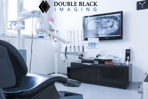
  • Viewing high contrast images in a dark environment multiple hours per day
  • Utilizing speech mics with Speech recognition software
  • In certain cases, performing procedures
  • Reporting test results
  • Preparation of lectures and presentations
  • More commonly, these tasks are being performed at home or from teleradiology workstations.

This is just a brief list of activities performed requiring repetitive motions.

Ergonomic workstations are designed to minimize the need for and impact of repetitive motions such as standing, bending, twisting, and leaning in performing your job.  Once these stressors are minimized, you can be more comfortable and productive when working.

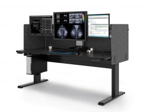
What does an ergonomic workstation look like?

Ergonomic equipment for workstations varies by profession.  As it relates to radiology workstations, ergonomic equipment includes:

  • Desks/tables that have:
    • Sit to stand adjustment capability
    • Ability to change monitor height independent of worksurface
    • Ability to easily and quickly change focal depth of monitors
    • Large usable worksurface to provide space for radiologist’s equipment: multiple keyboards and mice, voice recognition mic, trackball, etc. . . Ideally a single surface to maximize useable space in a given footprint
    • Ability to be quickly and easily adjusted/set up for each radiologist using the station
    • Dimmable back lighting to reduce eye strain
    • Electric sit to stand workstations/desks
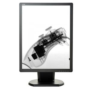
  • Monitors that:
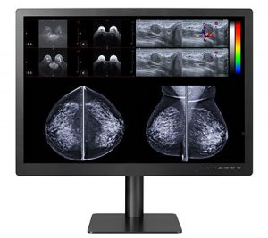
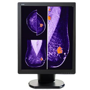
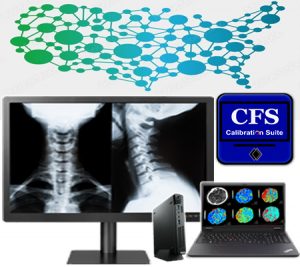
    • Meet ACR and FDA standards for brightness and resolution, ensuring images are DICOM Calibrated for proper diagnosis
    • Include productivity tools with the ability to dim worklist panels, limiting ambient light causing eye strain.  Cursor wrap functions eliminating continuous scrolling over the worksurface
    • Maximize viewing clarity from various angles
    • Reduce stress by offering adjustable height and angle
  • Chairs that provide
    • Vertebral support
    • Head support

What should you consider before implementing an ergonomic approach at your radiology practice?

Implementing an ergonomic approach is more than buying equipment that has a list of features.  It is important to see how specific features impact your patient care, medical staff, and overall satisfaction of the team.

A comprehensive ergonomic workflow rollout address every aspect of the practice and includes proper training on the use of the equipment as well as how can prevent injuries.

Technicians in radiology can benefit from an ergonomic approach as well.  Important areas to consider for these team members includes:

  • Manipulation of equipment necessary for performing procedures
  • Transportation of the equipment
  • Sit/stand desks for MRI and CT technicians
  • Positioning of patients for completion of the procedure
  • Ergonomic seating

How Does an Ergonomic Workstation Help Your Practice?

Ergonomics offers more than physical benefits. It results in increased productivity and efficiency, increased accuracy of reporting, a comfortable work environment, improved employee retention, and improved patient care.

The Association for Talent Development1 indicated that there are several factors that are improved when you focus on health and wellness:

  • absenteeism
  • retention and turnover
  • productivity
  • employee engagement

The costs associated with these factors is not small.

When an employee is injured, employers pay for more than the worker’s medical treatment. In its “NSC Injury Facts 2017,” the National Safety Council (NSC) calculates the average cost of an injury at $31,0002.

Radiation therapists appear to be at considerable risk for work-related musculoskeletal injuries, which corresponds with findings of studies on health care employees. They are reported to have a high prevalence of injuries (76%)3.  In fact, 58% of radiologists report symptoms of repetitive stress injury (many spend over 8 hours per day at their desks).

The most common site-specific injury is the lumbar back, followed by the neck and shoulders3. Other injuries can include repetitive stress injuries, visual problems, and headaches.

Productivity and Efficiency

When an ergonomic approach is used, staff can avoid bending, twisting, and awkward positions that cause fatigue, soreness, and lost time, as well as decreased comfort as the workday progresses.

Employees can be happier and have more energy when they can be comfortable when they work.  It’s easy to see that more energy put into helping patients results in patients who are happier with the care they receive.

Accurate Data Entry: A Surprising Benefit

Initially you might not consider that an ergonomic approach to Radiology workstations could impact the accuracy of data entry.  However, consider that you would have a higher level of fatigue throughout the day when you have a high level of stress on your body. Errors are more likely due to the higher level of stress and fatigue that results in decreased focus.

Ergonomic equipment increases safety and comfort throughout the day, meaning that employees are safer, more comfortable, and less fatigued.

A Comfortable Radiology Workstation adds Value and Savings

When care providers can adjust table height, view records through an optimally sized and correctly positioned monitor, and move easily around the facility/unit, they are more comfortable and experience less fatigue. This means that strain on backs, wrists, and necks is drastically reduced, or eliminated.  Ergonomic workstations designed to the unique demands of a job helps the radiology team do their job effectively and efficiently in comfort.

A ROI Comes in Many Forms

By now you realize that implementing an ergonomic approach requires investment of time and money.

Considering the costs of worker’s compensation, rehabilitation, and training of new radiologists, it is easy to see how you can quickly realize a return on your investment.

The biggest savings are seen in:

  • Less time off for injury
  • Fewer staff injuries and worker’s compensation claims
  • Greater accuracy in reporting and patient care
  • Healthier and happier staff with less turnover

When preventable injuries, burnout, and staff error are avoided, money typically spent on these items can now be spent where it is needed most.

Improved Patient Care is the Ultimate Reward

Imagine the improved patient care and improved patient satisfaction when:

  • Procedures are completed once because the staff could effectively and efficiently do it correctly the first time
  • Appropriate care is provided because errors are not made that might be related to fatigue or limited focus
  • Patients are more confident in the services they receive because staff treating them are more positive and energetic when providing care

These are just a few ways that an ergonomic workstation can improve patient care.

Why You Should Work with Double Black Imaging for Your Radiology Workstations

At Double Black Imaging, we don’t just sell you equipment to meet a standard ergonomic need.  We complete an onsite radiology workstation survey to assess what ergonomic equipment will be best for you and your radiologists.

We make specific recommendations for each surveyed workspace.  Our recommendations for your specific needs are based on our insider knowledge of PACS, mammography/tomography, clinical and surgical applications.

Contact us here, email us, or call us at (877) 852-2870, to arrange your Onsite Radiology Equipment Survey and determine what workstation will be best for you and your radiologists.


References:

  1. ATD Releases 2016 State of the Industry Report | ATD, January 9, 2023
  2. Employee Injuries: What Is the Real Cost to Employers? (workplacetesting.com), January 9, 2023
  3. Prevalence of Musculoskeletal Work-related Injuries Among Radiation Therapists – PubMed (nih.gov), January 9, 2023 Hanania AN, Cook A, Threadgill MP, Conway SH, Ludwig M. Prevalence of Musculoskeletal Work-related Injuries Among Radiation Therapists. Radiol Technol. 2020 May;91(5):414-421. PMID: 32381659.
17 Aug 2022

Comparisons of LCD Color and Monochrome Displays in Diagnostic Imaging

The types and number of imaging data handled by hospitals are increasing as we witness improvements in medical technology.  This is having an impact on the type of imaging you need to use, as well as the type of display you need to view and analyze images.  As a result, you will see combined use of monochrome and color images to achieve the greatest diagnostic accuracy.

The choice between LCD Color and monochrome displays in diagnostic imaging is not a simple one.  Each has some advantages over the other, and a comparison that considers the unique needs of your facility is warranted.

For example, both color and monochrome displays can use grayscale to view images.  The grayscale that is used can impact the quality of the image and the accuracy of your diagnosis.

Which display you choose can determine how you use grayscale to view your images, whether you can switch from monochrome to color images, and whether images are converted manually by you, or through automated analysis.

Not only will these considerations influence the accuracy of your diagnoses, but the amount of time invested in achieving those outcomes.  Display functionality can save you time or cost you time, depending on your priorities and choices.

Comparing LCD color and monochrome displays for radiology is an important consideration when choosing the type of display to use in your hospital.


What Is An LCD Color Display


LCD stands for Liquid Crystal Display.  Today most monitors that radiologists use for reading studies are LCD.  These displays look like flat panel televisions and computer monitors; however, medical-grade displays meet a higher level of standards, have greater functionality for medical imaging, and are more robust for your needs.

Most LCD monitors use Light-emitting diodes or LEDs as the backlight source.  This is a change from past monitors, which used a Cathode Fluorescent Lamp (CCFL) technology.  LEDs are slimmer than CCFLs, and this allows the display to be slimmer than those with CCFL tubes.

Another important consideration is safety.  CCFL technology uses mercury which is dangerous to the patient, health care workers, and the environment.

LED technology is a mercury-free source of light.  While LED contains other dangerous metals, it doesn’t present the same health and environmental risk of mercury.  LED and LCD life expectancy is greater than the old CCFL.


What Is A Monochrome Display?


Traditionally, Monochrome medical-grade displays have been recommended in diagnostic radiology because they have a higher luminance.  The higher luminance of monochrome displays makes it easier to see the entire grayscale in an image.

More specifically, monochrome displays have only one color of phosphor.  Looking at the name, you can see that “Mono” means one and “chrome” means color.  Everything (text and graphics) is displayed in that color.

If you perform a lot of imaging modalities such as chest CT, DR, and other imaging modalities that require higher brightness and contrast performance, then monochrome displays are a great solution for you.

Monochrome displays are significantly more expensive than LCD color displays.  Less expensive monochrome displays are available.  However, they come at a cost of reduced luminance which impacts image quality.  Decreased image quality is a significant compromise when considering the impact on diagnostic accuracy.


How Are They Similar?


Both monitors require calibration to maintain accurate functioning.  Medical grade displays are usually equipped with controls to facilitate grayscale calibration.  Monitors need to maintain calibration to the DICOM standard.  Maintaining calibration is particularly important when images need to be compared to previous images of the same person and condition on a different display.

Both LCD Color and Monochrome displays should be medical grade because they are designed for high volume use and facilitate viewing radiological images with the detail needed for accurate diagnosis and efficient completion of the analysis.

There can be some variability in uses that are recommended; however, based on the general characteristics of LCD Color and Monochrome displays are recommended for several of the same modalities.

For example, looking at LCD color and monochrome displays used for mammography and tomography, both are recommended for Computed Radiology (CR), Computed Tomography (CT), Digital Radiography (DR), Ultrasound, PACS and Mammography.

They come in similar sizes with similar numbers of pixels and resolution.  The viewing angle for these displays is not significantly different.  There is no difference in palette, video inputs, dimensions, and weight, and both have external power supplies.


How Are They Different?


Monochrome displays have a much higher cost. The trade-off is the longer life span of the display.  Also, they have higher luminance which facilitates efficiency and accuracy.

Color monitors have a lower maximum luminance compared to monochrome displays.  This doesn’t necessarily mean that diagnostic accuracy will be decreased.  The study by Geijer, et al.1, indicated that accuracy with color LCD and Monochrome monitors was not significantly different, but that the dwell time for color monitors was greater.

All displays can be windowed.  However, color displays often need to be windowed to improve image quality for specific area you are analyzing.  While a normal part of reading a digital image, it is more necessary in viewing on color displays, and this can reduce efficiency and limit the ability to compare objects in the window with those restricted from view.

Color and monochrome images should be viewed using different grayscales.  Monochrome images are best viewed using the DICOM 14 Grayscale Standard Display Function (GSDF) while color images are best viewed using Gamma 2.2.

In addition to modalities recommended for both displays above, LCD color displays are recommended for Nuclear Medicine, 3D, Positron Emission Tomography (PET), and Tomosynthesis.  LCD Color displays consume more power than monochrome.

An advantage of color displays is that they can show color information.  As medical technology advances and more modalities use color, this is an important consideration.  Modalities that already use color include Doppler ultrasound, 3D reconstructions in computed tomography (CT), functional magnetic resonance (MR) imaging, and nuclear medicine, including PET.

Given that color displays are significantly less expensive, they can be replaced up to four times more often within a fixed budget1.

Meanwhile, monochrome displays have a higher contrast ratio and brightness than LCD Color displays.


Selecting The Right Display For Your Office


Displays now function to read images and adjust between color and monochrome, or to allow easy switching from one functionality to another.  This makes the less expensive, LCD Color displays more attractive.  However, there are circumstances when the monochrome display is the better choice.

Which display you choose will be influenced not only by cost, but also by the current and future needs of your hospital, which is determined by the types and numbers of modalities you perform as well as what you anticipate you might add to the modalities you perform during the life of the display.


Get Your Radiology Displays From Double Black Imaging


Double Black Imaging understands the importance of displaying high quality images while containing costs.  We can help you choose the best display options for your facility and your budget.  We offer a range of options that are the best LCD Color and Monochrome displays on the market.  Contact us today to set up a discussion about how we can help you with your next display purchase.


1 Geijer H, Geijer M, Forsberg L, Kheddache S, Sund P. Comparison of color LCD and medical-grade monochrome LCD displays in diagnostic radiology. J Digit Imaging. 2007 Jun;20(2):114-21. doi: 10.1007/s10278-007-9028-5. PMID: 17340227; PMCID: PMC3043910.

12 May 2022

What Are Surgical Displays And Monitors?


Surgery monitors and ER monitors provide high quality images that facilitate your ability to successfully perform surgeries and procedures. A quality monitor allows you to see the patient’s anatomy in detail and make diagnostic and treatment decisions for the best patient outcomes.

Medical-grade monitors are used in operating rooms, emergency rooms and medical offices.  Displays you use in image-guided procedures such as endoscopy (gastrointestinal, arthroscopic, laparoscopic, or robotic guided surgery) and interventional radiology are usually referred to as surgical displays or surgical monitors.  You can use them to display a variety of critical information, including patient data, x-rays, vital signs, brain activity, endoscopy images, PACS images, surgical feeds, etc.


Types of Surgical Displays


Surgical displays come in a variety of forms, shapes, and sizes.  These devices include displays you can use near a patient (near-patient displays), large screen operating room displays, user interfaces (touch displays), and control displays which are often used in interventional radiology.

Surgical displays can offer high-definition or 4K visualization for presented visual images.  These displays can be mounted on your radiology practices wall, on an equipment column, or on a surgical light arm.


Diagnostic Displays


As a physician or radiologist, use a diagnostic display to view various diagnostic images like Computerized Tomography (CT) scans, Magnetic Resonance Imaging (MRI), and X-rays.

Medical facilities are relying on diagnostic displays more as new procedures require you to employ endoscopic visualization to perform surgery or diagnostic procedures.

High Definition and Ultra High-Definition Surgical Displays


As you rely more on image-guided surgical devices, you can appreciate the movement from High Definition to Ultra High Definition (4K) technology incorporated into new systems and monitors.  A 4K monitor provides four times the image detail compared to High-Definition images. This means that you can see more anatomical detail and more clinically relevant information in order to provide the best service to your patient.

Another benefit of 4K displays is that they provide a wider color gamut.  It provides you with a broader spectrum of reds – which is especially helpful in the OR.  It also provides more detail and color-corrects images, which means you can differentiate tissue types and structures such as nerves, blood vessels, organs, and organic material within your patient.

It is also important to note that with the detail gained with 4K resolution, you gain a better holistic perception and a more immersive environment to operate with greater precision.


Large Format Displays


Simply stated, Large Format Operating Room displays give you a larger viewing area.  They can be mounted on the OR wall so that your entire team can view the surgical site during the procedure.

This feature makes large format displays ideal for use in your hybrid ORs, interventional X-ray and cardiovascular suites.


Important factors in a Surgical Display


           Crisp Image

Clear, crisp images mean you won’t have artifacts and visible pixels.  So, you can see the fine detail of the surgical site.  Some surgical displays offer multi-image visualization for viewing multiple images simultaneously in high quality.

The backlight stabilization of surgical displays ensures consistent image brightness and maintained focus for moving images.

Finally, you can see details clearly, even when you are not standing directly in front of the display.  So, there is no need for frequent readjustments of display location and angle.

          Fan-less Design

A fan-less monitor has the advantage of being easier to clean and disinfect, preventing the spread of contaminants and saving your staff time and resources.

Also, the fan noise is eliminated, enabling your team to communicate more effectively, and reducing concerns about errors related to noise interference.

          Ventless Design

Surgical monitors with vents require additional time and effort to clean. You risk contamination of your hands, the patient, or surfaces near the patient if the pathogens are not completely removed by thoroughly cleaning all surfaces.

A ventless design has two great advantages:

  1. It reduces the surface area that you must clean, making cleaning easier and less time consuming.
  2. It also reduces areas where dust can settle, and bacteria can grow.


          Impact-resistant Design

Surgical monitors with impact-resistant shields protect the panel from physical damage, which means your technology investment is protected against collisions with lights, IV poles, and other structures in the OR.

This is particularly important when your staff is working quickly to accommodate frequent turnovers for new procedures.

         Medical Grade Power Supply

A Medical grade power supply has a long lifecycle and minimizes the risk of shock.  Also, you gain protection against a technical or power failure so you can maintain critical information during a procedure.


Why are Double Black Imaging Surgical Displays the Best?


Double Black Imaging is committed to providing you medical imaging technology that enhances your diagnostic capabilities.

Here are just a few of the many reasons Double Black Imaging Displays are the best:

  1. Our displays provide bold, crisp images even when you view them from a distance.
  2. We provide you with the world’s most technologically advanced Surgical Displays.
  3. They are built to withstand years of use.
  4. Our monitors give you unmatched quality, clarity, and consistency.
  5. We offer a full line of splash-proof Surgical Operating Room Displays.
  6. Our solutions meet and exceed all surgical imaging requirements.
  7. Our displays offer full HD resolution, ultra-high brightness, and
  8. They support a wide variety of inputs so you can interface with your new or technologies and eliminate costly signal conversion, signal degradation and image lag.
  9. Our monitors to deliver exceptionally fast response time when viewing live video or motion, with in versatility for picture in a picture or picture by picture format.
  10. We guarantee that every display has certification for classification as a medical display.


Why Choose Double Black Imaging?


At Double Black Imaging, we base each product and business decision on how to best serve your needs.  We strive to build a long-term relationship with you by offering the most advanced equipment at competitive prices.

Double Black Imaging began in 2002 with the goal of making medical imaging more efficient and improving healthcare.  We maintain that standard by providing the best value in the industry and by offering technologically advanced medical imaging solutions that enhance diagnostic capabilities, along with exceptional customer service and support.


Premier Customer Service


Double Black Imaging’s commitment to making the best decision for you, our customer goes well beyond the sale of OR Monitors.

Surgical Displays and Monitors


Our industry-leading customer service means we provide you the necessary knowledge and support, before and after your purchase.

To learn more about your ideal OR Monitors or our other products and services, contact us today at (877) 852-2870 or email us at sales@doubleblackimaging.com.

25 Mar 2022

No Surprises Act in Radiology

No Surprises Act in Radiology: A Summary of Key Factors

What is the No Surprises Act in Radiology?


When treated by facilities or physicians outside their insurance network  patients could receive large, unexpected bills. This is known as surprise billing.

Simply stated, these providers do not participate in an insurance plan and typically bill and are reimbursed at a different rate.

The No Surprises Act is a federal law that limits the financial obligations of patients to out-of-network providers. This applies to emergency and non-emergency situations.

No Surprises Act billing rules state that a patient’s out-of-pocket expenses should be the same for out-of-network providers as it is for providers in-network. Depending on the patient’s insurance plan, this will be the co-payment, co-insurance, or deductible that is normally owed to in-network providers.


When was it voted on?


In December 2020, Congress signed the Consolidated Appropriations Act.  The No Surprise Act is part of that legislation.


When did it go into place?


The No Surprises Act effective date was January 1, 2022, so you are now expected to follow the law.


What has changed because of this act?


If your state has already enacted no surprise billing legislation, the No Surprises Act will not supersede it.

For states that have not yet enacted a surprise billing law, the No Surprises Act will result in several changes.

For non-emergency care, you will need to obtain consent prior to providing services.

Secondly, the patient cannot be billed directly.  You will need to bill and negotiate reimbursement through the insurance provider.

Once you have received payment, you have 30 days to negotiate or appeal the amount.

Finally, you will need to use the established system to negotiate and/or request arbitration of reimbursement rates.


Services Covered


If a patient requires emergency care, you should provide services until they are stabilized.  As an out-of-network facility, you must then address the issue with the patient to obtain consent to be transferred to an in-network facility or physician.

The patient can also consent to continue receiving out-of-network services from you.

Patients who sign consent waivers will then be responsible for the balance of uncovered expenses.

In a non-emergency situation, if your facility has in-network providers, you are permitted to request a consent waiver.  This allows you as the provider to balance bill the patient.

In these non-emergency situations, it is important to note that you need to give the patient a written consent waiver 72 hours prior to the procedure/medical service.

You need to include the following in the consent waiver:

  1. A declaration that you are not in the patient’s insurance network
  2. An estimate of the cost of your services that is as accurate as possible
  3. If your facility is in-network, a list of in-network providers at your facility
  4. A summary of any prior authorization or care management requirements that must be in place prior to the procedure/service
  5. A statement that the patient can opt to receive care from an in-network provider

When the non-emergency procedure will occur in less than 72 hours, you must provide the notice/consent waiver at the time the appointment is made.


Independent Dispute Resolution (IDR)


The No Surprises Act provides a system for negotiating reimbursement.  Please note, you only have 30 days to come to a resolution with the payer or to file a dispute if you cannot come to an agreement.

If you fail to reach an agreement with the payer, you or the payer have four days to activate the Independent Dispute Resolution (IDR) system.

Once the IDR is initiated, you and the payer will each submit an amount to an arbitrator, who will select one of those amounts as the settlement.

You cannot split the difference or request further negotiation.

Once decided, neither you nor the payer can repeat the process for the same service or procedure with each other for 90 days.

The losing party pays the cost of the arbitration process.

Over time, you will learn what level of payment is acceptable to the arbitrator, and this should limit the need for IDR appeals.


Impact on Radiologists


Radiologists and other ancillary service providers are not able to use consent waivers. This means you must abide by the in-network payment policies of the Act.

The No Surprises Act does not exempt patients from financial responsibility. You can expect them to pay what they would pay if you were in-network. This will be clearly communicated to you on the Explanation of Benefits (EOB) that the insurance company sends with their payment to your practice. You can bill the patient for this amount once you have accepted the payment or a payment has been negotiated. You can bill them for their co-payment, co-insurance, or deductible as indicated in their insurance plan.

Time and resources spent negotiating rates and engaging in arbitration, will impact finances for you and the payer. It is in your best interest to establish a system to minimize this impact.

One solution is to try to reach an agreement with out of network (OON) payers. This will eliminate potential disputes about reimbursement, special claim handling and costs of arbitration via IDR, so it will be beneficial to them to make this agreement with you.


Importance of developing an internal process


You will learn about the charges and reimbursement for OON patients as you settle disagreements with the payer.  However, handling charges for these patients will require a different system than what you use for in-network billing.

Be sure in-network vs. out-of-network status is clearly determined from the beginning for these patients so their reimbursement claims can be processed correctly.

Even though patients are out-of-network, you should submit your initial claim to the insurance company for your full fee.

Your system should tag accounts so that you can review payments when they are received.  This will avoid billing the patient for the balance rather than the allowed amounts.

You also want your staff to determine whether the payment is acceptable to your practice within the 30-day negotiation window.  This way, you don’t miss the opportunity to negotiate a rate or file a dispute.

Your staff can respond quickly and know if negotiation is necessary. If you take the time to create a list of reimbursement rates that you find acceptable for your procedures. Also, over time you will learn from the arbitrator what is an acceptable reimbursement level.

Claim processing can be more efficient if you create a fee schedule within your accounting system.


About Double Black Imaging


Learn More About No Surprises Act in Radiology

Since 2002, Double Black Imaging has brought high quality computing and imaging products to the market in response to changes in the market technology and medical care. We offer solutions that will help you implement these changes effectively and with minimal effort.

Contact us today at (877) 852-2870 or email us at sales@doubleblackimaging.com to learn more about how our products can help you comply with these new requirements without a negative impact on your budget.