Category: Radiology & Diagnostics

15 Jun 2021

Filling the Gaps in Radiology Workflow

Filling the Gaps in Radiology Workflow

Load balancing, or equitable distribution of radiology workflow, has been a longstanding and widely disputed issue in radiology stemming back to the pre-PACS era. Unequal workloads resulting in unequal compensation have threatened to divide many imaging practices. The introduction of PACS solved many of the imaging and reading problems radiologists used to face, and even saw some groups doubling production, but did not fill in all the gaps in radiology workflow.

The advent of PACS also occurred during a period of healthcare reform that demanded higher production and higher quality benchmarks while lowering reimbursements. And new problems emerged as easier cases were quickly processed while complex cases sat unattended on work lists for days. This has resulted in a full circle return to unequal workload distribution and conflict among radiologists.

Why Is Good Radiology Workflow Important?

Balanced medical imaging workflow is an elusive goal radiologists have pursued for decades due to its widespread effects on both the profession and the healthcare system. And while a radiology practice or department completes many tasks, a study has shown that roughly 80% of radiology workflow problems can be attributed to 20% of its cases. Identifying these problems can help bring about customized solutions.

With radiology being a $100-billion dollar industry, leaders in the field point out “approximately $15 billion to $20 billion of radiology’s expenditures are unnecessary and avoidable. Either the right test is conducted at the wrong time, or patients receive a repeat exam. Regardless, improving workflow can save not only money, but it can also save practices’ and departments’ time.”

Besides saving money, more efficient workflows can help reduce delays in patient care as well as medical record and clinical errors. This can be accomplished through real-time communication to manage and transfer patient data.

Another gap in radiology workflow as a result of the evolution of imaging technology is communication between radiologists and clinicians. It used to be that clinicians had to call and speak to the radiologist about imaging results or physically walk over to the radiology department to discuss and view patient results. Since the introduction of PACS, clinicians can review radiological reports and images anytime from any location. While the importance of face-to-face communication may be debatable, the importance of accurate and timely communication between radiologists and clinicians for patient safety and care remains important to all physicians.

Improved imaging and workflow efficiency also results in better patient care as a result of improved consistency, higher quality imaging, and individualized medical care.

Better working relationships with care providers and more effective use of office hours are other important benefits when radiology workflow is handled efficiently and fairly distributed.

How Has Technology Changed in Radiology?

It is now common to see health systems with different PACS installations from different vendors spread across the networks of imaging centers and hospitals. A shared goal in these systems is to aim for equitable case distribution amongst radiologists across these vast networks, and to try to improve efficiency and workload balance while ensuring the most qualified radiologists are assigned the most complex cases.

Brady concurs, explaining “Orchestrating workflow more efficiently is a win-win-win for patients, physicians, and staff. Patients benefit when the radiologist who’s best equipped for and experienced in reading that type of subspecialized exam is automatically chosen to read it, and it’s routed quickly and efficiently.”

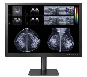
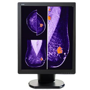
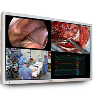
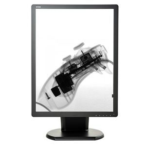
One of the medical imaging workflow problems Double Black Imaging solved is requiring different displays depending on the modality being read. It is not conducive to a smooth workflow if a radiologist has to change workstations to read CT,MR, Nuc Med, Chest, Bone or Mammo/Tomo. Their 8MP and 12MP large format displays enable doctors to read any modality from one large screen. 5MP Color displays provide radiologists a dual monitor set up for multi-modality imaging if two screens are preferred over one.

How Has Technology Changed Medical Imaging Workflow?

To help improve communication between radiologists and clinicians, some radiology departments have found unique ways to use imaging technology. For example, NYU Langone Medical Center in New York City has incorporated virtual radiology rounds and multimedia reporting to improve communication with clinicians.

For virtual radiology rounds, clinicians arrange online meetings with radiologists to review images and reports. “The clinicians and radiologists then use web cameras to see each other during the meetings, and they also remotely share a computer screen and mouse. This allows both parties to point out details on the images, provide feedback, and ask questions — just as they would during in-person consultations….” Not only has this improved communication with clinicians, but radiologists also feel it makes them more “visible” and helps create greater value for patients and referring physicians.

Due to the success of these virtual rounds, radiologists at NYU Langone plan to begin offering virtual consults on demand to physicians’ offices at hospital outpatient departments as well. When the clinician loads the images into the hospital’s PACS, a message will pop up asking them if they want to speak to a radiologist. If the clinician clicks ‘yes’, the radiologist assigned to those cases receives a message on their phone or other electronic device advising them an on-demand consult is being requested. Radiologists at NYU Langone anticipate this service will provide significant value to both physicians and patients while restoring the role of radiologists as consultants and integral members of the clinical care team.

What Impact Will AI and Machine Learning Have on Radiology?

Artificial intelligence, also known as machine intelligence, refers to a wide scope of intelligent functions that can be performed by computers such as planning, language processing, knowledge representation, problem solving, and actual “learning” based on previous data it has reviewed and processed.

While AI is not expected to replace radiologists, it has been compared to the autopilot function on airplanes that relieves pilots of performing repetitive and tedious but necessary safety checks and tasks. Arazi notes that once the COVID-19 pandemic tapers off, AI will play a key role in helping radiologists save lives post-pandemic. He suggests it will do this by helping prioritize growing numbers of non-urgent but important procedures that were set aside due to the pandemic, such as cardiovascular imaging, bone-health scans, and mammography screening.

AI also has the potential to change the role of radiologists from active disease diagnosticians to proactive diagnosticians, assisting patients with early intervention and even preventive care by identifying potential problems before they occur.

Similarly, machine learning is one type of artificial intelligence that can be used to pull, organize, and analyze information from big data sets created by large electronic medical record systems. This information can then be used as a basis for anticipating patient outcomes and clinical decision-making.

In the future, machine learning may also help improve different components of radiology workflow such as radiology reporting, postprocessing and dose estimation, triaging, order scheduling, quality control of exams, detection and interpretation of findings, and support systems for clinical decision-making.

For all your radiology imaging needs, including innovative software solutions and superior medical-grade displays, contact our diagnostic imaging experts at Double Black Imaging. Our unparalleled customer service and highly knowledgeable staff set us apart from the competition, making us the first choice for all your imaging needs.

Source List:

https://www.radiologytoday.net/archive/rt0220p8.shtml
https://www.jacr.org/article/S1546-1440(18)30257-6/fulltext
https://www.diagnosticimaging.com/view/how-streamline-radiology-workflow
https://www.ncbi.nlm.nih.gov/pmc/articles/PMC6650448/
https://www.acr.org/Practice-Management-Quality-Informatics/Imaging-3/Case-Studies/Information-Technology/Closing-the-Gap
https://www.ncbi.nlm.nih.gov/pmc/articles/PMC6542626/
https://www.weforum.org/agenda/2020/10/how-ai-will-change-how-radiologists-work/

15 May 2021

Ensuring and Measuring the Value of Radiology Imaging

Radiology Imaging Value

Showing radiology imaging value is easier said than done. That is because the term “value” has different meanings to the different stakeholders involved. Insurance providers and Medicare see value in diagnostic testing when patient results come back positive for a health condition, particularly if the condition is caught early and leads to reduced healthcare costs down the road.

For hospitals and clinics, measuring radiology imaging value includes the ability to obtain high-quality reliable images that reduce the amount of physician time required to make a diagnosis.

For a patient, the value in radiology imaging is in its ability to potentially save their life from a serious health threat, or to help identify a course of treatment so they can begin to feel better again.

So the question becomes which stakeholder’s definition of value is most important? And once this is determined, how can this “value” be measured?

The Complexities of Showing Value in Radiology Imaging

As Kruskal and Larson point out “The question is not whether radiology will survive, but rather what our role will be in the specialty and in the medical field going forward.” Because the term “value” is tied to healthcare costs for payers, radiologists face a number of potential changes to their profession that may not be of their choosing and which they may have little control over.

While technological advancements have resulted in improved capability to diagnose certain health conditions, as well as the ability to diagnose with improved accuracy, they have also driven up the cost of delivering these services. This has caused insurers and others to question the value some of these services offer.

With healthcare costs becoming an increasing financial burden for households and society as a whole, diagnostic imaging has become the scapegoat to blame for these ever escalating costs. In response to political pressure, payers are actively looking for ways to reduce these soaring expenses. Insurers see medical imaging as one area where they can significantly reduce healthcare costs going forward.

Different Types of Metrics

Due to the complex nature of radiology, several different types of metrics are used to monitor and measure performance. Some of the most common metrics in radiology include customer satisfaction, regulatory compliance, clinical productivity, academic productivity, and financial performance.

The key to selecting appropriate metrics that will actually measure what needs to be measured is in choosing indicators that closely align with an organization’s mission. When specific metrics are chosen keeping an organization’s mission in mind, improved service delivery and performance is more likely to be achieved.


  • Process Metrics

    Metrics that measure and provide an overview of the state of an organization’s processes or operations are known as process metrics. Some examples include:

    • Report turnaround time
    • Patient access time
    • Percentage of calls answered within a specified time frame
    • Percentage of biopsy results delivered to the primary care physician and the patient
    • Percentage of undictated cases at a specific number of days
    • Percentage of carotid imaging reports with distal internal carotid artery size as diameter
  •  


  • Outcome Metrics

    Outcome metrics measure the outcomes or clinical impact of an organization’s processes. Examples include measuring the percentage of:

    • Complications
    • Adequate biopsy tissue
    • Falls incidents in the radiology department
    • EVAR patients without major complications
    • Examinations with contrast media reactions
    • Patients with CVC insertion using sterile barrier technique
  •  


  • Volume-Based Metrics

    Radiology practices have historically used volume-based metrics to monitor their performance in patient care delivery in categories like efficiency, customer service, financial performance, and staff productivity. This includes:

    • Staffing efficiency
    • Report turnaround time
    • Equipment use and downtime rates
    • Patient access and wait times
  •  


  • PPACA-Based Metrics

    Since the Supreme Court decision to support passage of the Patient Protection and Affordable Care Act (PPACA), several new radiology metrics have been introduced.

    For example, the federal government has introduced initiatives such as the Physician Quality Reporting System (PQRS) that encourages physicians to report quality measures that may result in financial penalties.

    In an attempt to control the rising cost of insurance premiums, insurers have implemented programs and contracts aimed at improving accountability and patient outcomes while reducing service costs.

    With the integration of hospitals and healthcare systems, radiology practices that contract with these systems are required to be accountable for some new performance metrics as well. These new metrics are found in categories such as subspecialty expertise, critical findings, turnaround time, use of voice recognition, peer review, and extended hours.

    National radiology professional societies such as the American College of Radiology (ACR) and the American Board of Radiology (ABR) are also working to develop metrics that radiology groups and departments must meet to receive modality and site accreditation or the Maintenance of Certification.

  •  


  • Value-Based Metrics

    More recent value-based metrics measure health outcomes achieved for every dollar spent. This is done by categorizing outcomes at the patient level that are disease specific and based on an overall “episode of care”, regardless of how complex a health episode may be. An episode of care is measured from the onset of symptoms to cure or death of the patient.


How Radiologists Can Improve Value-Based Metrics


Radiologists can begin to make the shift towards showing radiology imaging value by implementing the following steps:

  1. 1. Commit to continuous learning. Colleagues and other facilities and practices can be a rich resource of information if they have found better ways to work more efficiently and cost-effectively. Be willing to reciprocate and share your knowledge with others.


  1. 2. Understand the needs of your referring physicians. Find out what they would like to see improve or change about how you work and then figure out ways to make this happen.


  1. 3. Communicate Effectively. Make a point of calling your referring physicians when needed to discuss a patient’s care. Ensure reports are free of errors and unclear interpretations and are produced in a timely manner according to national guidelines.


  1. 4. Focus on teamwork. Build trusting collegial relationships with referring physicians to help improve the patient’s’ healthcare experience.


  1. 5. Champion the physician role. Radiologists are important stakeholders in the value equation. Offer to join committees and hospital boards and respond positively to feedback.


  1. 6. Critically reflect on how to improve your practice. Be willing to explore new ideas and systems that improve patient outcomes and workflows while reducing costs.


  1. 7. Collaborate with your IT department to improve workflows. Look for ways to put the needed systems in place to make it easier and faster to produce, read, share, and save reports and communicate about patients.


  1. 8. Proactively look for ways to use resources more efficiently. Identify ways to reduce repeat imaging, inappropriate recommendations, and safety related incidents. Look for ways to increase turnaround time and reduce negative patient experiences and feedback. Also, look for ways to demonstrate how the consultative and coordination of services roles in radiology reduce costs through saved physician time “downstream” in a patient episode.

 
Showing and measuring radiology imaging value may not be easy. However, it is essential moving forward as insurers and hospitals increasingly use value-based metrics as performance indicators tied to healthcare reimbursement.

At Double Black Imaging, we are committed to providing innovative imaging and workflow solutions. Our goal is to help providers significantly improve diagnostic imaging quality and stability while improving efficiency and reducing healthcare costs. Contact us to discuss ways to improve these performance metrics in your practice today.

Source List:

https://www.sciencedirect.com/science/article/abs/pii/S1546144018314595
https://www.radiologytoday.net/archive/rt0518p16.shtml
https://www.radiologybusiness.com/topics/quality/survey-radiology-quality-metrics-practices
https://pubs.rsna.org/doi/full/10.1148/rg.2015140221#tbl4
https://www.medicaltranscriptionservicecompany.com/aligning-radiology-metrics-with-the-goals-of-value-based-care/
https://www.diagnosticimaging.com/view/how-value-based-care-affecting-radiology
https://insightsimaging.springeropen.com/articles/10.1186/s13244-020-00941-z
https://pubmed.ncbi.nlm.nih.gov/23025865/

20 Apr 2021

Prioritizing MRI Safety with Safe Imaging

The evolution of MRI safety has become more challenging than ever for radiologists, MR technologists, and referring physicians.

As Sammet explains” The increasing clinical demand for Magnetic Resonance Imaging (MRI) with its superior soft-tissue contrast compared to other radiological imaging modalities and potential physiological and functional applications has contributed to the installation of almost 30,000 MRI scanners worldwide.”

Combined with this is the rapid increase in the number of patients who have implants due to advancements in medical implant technology.

This growth in clinical demand and the number of MRI scanners installed has required that significantly more health professionals be educated about MR safety to protect patients, themselves, and others who may be exposed to the MR environment.

What Is MRI and What is it Used For?

Magnetic Resonance Imaging (MRI) is a non-invasive imaging technology that produces detailed three-dimensional images of the body’s anatomy. The technology works by stimulating protons in the water of living tissues and then identifying how the protons’ rotational axis changes.

This is done by using extremely strong magnets that create a strong magnetic field forcing protons in the body to align with the magnetic field. By pulsing a radiofrequency current through the body, the protons are forced to spin out of balance as they strain against the pull of the magnetic field.

Then the radiofrequency is turned off allowing the protons to realign with the magnetic field which releases energy that the MRI sensors detect. How long it takes for the protons to realign with the magnetic field and the amount of energy released when this occurs differs depending on the chemical composition of the molecules and the environment. Using these measurements, physicians can identify the different types of body tissue.

Physicians use MRI to identify differences between healthy and unhealthy tissue in the body. It has become the preferred imaging procedure for diagnosing potential medical problems. Many body tissues can be assessed using MRI including the heart, blood vessels, internal organs, abdomen, pelvic area, brain, spine, breasts, and joints in the body.

How Safe is MR Technology?

Millions of MRIs are performed in the US each year. And according to the FDA, approximately 300 adverse events are reported from healthcare facilities, distributors, manufacturers, and patients related to these scans. Overall, this demonstrates the safety of MR technology for patients and healthcare personnel when the appropriate safety precautions are implemented and correct equipment is used.

Why Has MR Safety Become More Challenging and Complex?

MR technology has become increasingly complex not only because more scanners are being used and more procedures are being done, but also because scanners with higher field strengths are being developed and used in hospitals, institutions, and private practice.

In addition, radiologists are not required to have any formal training about MR imaging physics or MR safety concerns related to this form of imaging as Emanuel Kanal, MD, director of magnetic resonance services at the University of Pittsburgh explains.

The complexity and challenges related to MR safety are due primarily to three factors:


  1. The sophisticated technology that makes MR imaging possible


    The static magnetic field of an MRI machine is incredibly powerful. For example, a 1.5 T magnet exerts a magnetic force that is 21,000 times greater than the earth’s magnetic field. With this force, small metal objects such as hairpins and paper clips can be made airborne and move at speeds of up 40 mph into the machine, injuring a patient and anyone in the path of the flying object.

    Larger medical objects made of stainless steel such as scissors, hemostats, clamps, as well as oxygen tanks, metal gurneys and even floor buffing machines can become projectiles and injure people in the room as well.

    For this reason, all metal objects regardless of their size must be kept out and away from the room housing the MRI machine.

    Biological risks including cell growth and aberration and some reversible effects such as headaches, irritability, fatigue, and hypotension have been noted in human subjects exposed to static magnetic fields of 2.0 Tesla and above. Most MRI equipment currently produces fields less than 2.0 T which have not produced any known biological effects. However, the FDA recently approved the first 7 T MRI system for use in the US in October 2017 so these risks may now exist.

    One of the risks related to the radiofrequency (RF) field is the risk of current-induced thermal burns to the patient’s skin. This can occur if the RF field produces currents in wires that are near or attached to the patient. The RF field may also produce currents in intra-cardiac leads causing unintended cardiac pacing. Lengthy imaging procedures can also increase core body temperature although this is primarily a concern in infants.

    Cryogenic risk exists if the magnetic field is shut down accidentally or for planned maintenance. When this occurs, liquid helium inside the magnet begins to turn into a gas that may seep into the scanning room and displace oxygen leading to possible asphyxia of the occupants.


  2. Patient variably


    One of the greatest concerns about scanning patients is if they have implants, what material these implants are made of, and how these implants may react when exposed to the MR field. While the FDA allows manufacturers to market certain implants as “safe” for MR imaging “at or up to certain conditions under which they were tested”, MR environments are highly variable and may exceed these test parameters.

    According to Kanal, this variability may result due to different strengths of the MR energies at different positions in the MR scanner and scan room as well as at different positions throughout the patient’s body. And “How they vary can and do differ with different MR scanners and gradient coils and RF transmitting coils among varied scanners of the same or different MR manufacturers.”

    For these reasons, Kanal developed a smartphone app called MagnetVision that has all this information built in so it is able to calculate in real-time the effect of MRI on a particular implant, coil, MR scanner or patient.


  3. The increase in imaging procedures performed

    The rapid increase in MRI technology and software has also led to new imaging procedures being developed including multi-contrast imaging and lung imaging. With new procedures and technology come issues of safety that must be evaluated, documented, and communicated to those using this new technology with patients.

    When these potential MR safety issues are understood, proper safety protocols are followed, and the correct equipment is used, MRIs are safe and extremely effective in producing exceptional images that contribute to timely patient diagnosis and treatment.

    To meet our customers MR safety needs, Double Black Imaging offers two different sizes of MR Safe Displays that are fMRI compatible and provide a 2.3-megapixel color LED backlit display. They are available as 24-inch and 32-inch LCD displays to accommodate varying space constraints and bore sizes.

    Both LCD medical displays provide lag free output and no interference with the scanner even when positioned at the bore exit. Display specifications include an extraordinary spatial spatial uniformity of 2% over the central 75%, ultra-high brightness of 1000 cd/m2, and a contrast ratio of 1000:1 resulting in unparalleled MR image quality.

    At Double Black Imaging, we understand the challenges technicians and radiologists face in producing high quality images while keeping patient and healthcare personnel safety as their top priority. Contact us to learn how our MR Safe displays can help meet your practice’s imaging needs.


Source List:

https://www.ncbi.nlm.nih.gov/pmc/articles/PMC4848040/
https://www.itnonline.com/article/recent-advances-mri-technology
https://www.nibib.nih.gov/science-education/science-topics/magnetic-resonance-imaging-mri
https://www.ismrm.org/resources/information-for-patients/
https://www.mayoclinic.org/tests-procedures/mri/about/pac-20384768
https://www.fda.gov/radiation-emitting-products/mri-magnetic-resonance-imaging/benefits-and-risks
https://www.diagnosticimaging.com/view/qa-tackling-complexity-mr-safety
https://physicsworld.com/a/mri-safety-an-urgent-issue-for-an-increasing-crowd/
https://radiology.ucsf.edu/patient-care/patient-safety/mri/potential-hazards-risks
https://www.itnonline.com/content/fda-clears-first-7t-mri-system-magnetom-terra
https://www.itnonline.com/article/recent-advances-mri-technology

23 Mar 2021

WHY TRANSITION TO RADIOLOGY READING FROM HOME?

Radiology from Home

Since the widespread emergence of COVID-19 in North America in early 2020, remote radiology reading from home (also known as teleradiology) has seen a surge in popularity. Teleradiology refers to radiologists receiving images to interpret from a facility in a different geographical location than where the radiologist performs the reading. This may be in a country in a different time zone halfway around the world or in the radiologist’s home located just blocks from the hospital they are contracted with.

What’s the incentive to shift to remote radiology reading?

Social distancing requirements to help stop the spread of the virus during the pandemic has required a radical shift in how radiology is practiced in hospitals and healthcare centers.

Mohammed Imran Quraishi, MD, assistant professor of radiology at the University of Tennessee, and colleagues surveyed 290 geographically diverse institutions to determine how many sites were transitioning to teleradiology as a result of the pandemic. They received 174 responses and found a significant number of sites had radiologists installing workstations at home (65.2%) and a sizable movement to internal teleradiology (73.6%) to replace daytime shifts.

Beyond the safety related reasons for shifting to more remote radiology reading, performing radiology from home offers increasingly strong personal and economic incentives for physicians as well.

Jackson notes “When teleradiology first entered the industry as a career option, it was pigeon-holed into being an overnight service only. Radiology practices used teleradiology as a way to avoid overnight call. However, in recent years, reading images remotely has ballooned as a viable career option for radiologists who simply don’t want to work in hospitals or imaging centers.”

And if the increased flexibility of performing radiology from home is not enough to convince radiologists to consider working more at home both during and after the pandemic, the economic trends in the sector might be.

In February 2020, Grand View Research, an India & U.S. based market research and consulting company, reported the size of the global teleradiology market is expected to hit $10.9 billion USD by 2027. Growth in this sector is expected to be driven by a shortage of radiologists paired with an increased demand for imaging procedures.

The fastest growth is expected to occur in CT. This is due to quicker testing, widespread use, high-resolution image quality and the arrival of newer 4D generation systems.

In 2019, x-ray had the largest share of the teleradiology market due to its broad scope of application from identifying bone fractures to dental injuries. Increased demand for core diagnostics and imaging at the bedside combined with new technologies is predicted to drive growth even more.

    1. Flexible work
      Michael Yuz, MD, an executive radiologist with USARAD, a radiology-on-demand company points out, “As the opportunities have expanded, so have the positives that come with the work.” However, he is quick to point out that while radiologists working from home can choose their days and hours of work, they will not end up working less. In fact, he says it’s not unusual for radiologists working from home to work twice as many hours as their colleagues practicing in hospitals or imaging centers and to routinely put in 12-14 hour workdays.


    1. Better work-life balance
      Being able to choose what days and hours one works and the ability to work at home allows teleradiologists to have more freedom to decide where they want to live. It also allows them to spend more time with family and friends. This results in radiologists who are happier with where they work and live and as a result, they experience less work-related stress according to Michael Rothman, MD, a teleradiologist based in Bethlehem, PA. Quraishi et al. confirmed this in their study stating 64.8% of respondents who transitioned to working from home during the pandemic reported decreased stress levels. And this occurred while 96% of respondents found an improvement or no change in turnaround times for readings. Michael P. Recht, MD, Professor and Chairman of NYU Langone Department of Radiology “said he is ‘very much’ in favor of remote reading and noted that about one-quarter of his faculty took part in this practice—thanks to a wellness initiative—prior to COVID-19.” He says almost 70% of his radiologist staff have taken part in the program during the pandemic with these physicians experiencing improved work-life balance due to the increased autonomy and flexibility in their work.


    1. Ability to select a subspecialty
      As Yuz explains, most imaging facilities expect their radiologists to function as generalists. However, teleradiologists working from home have the ability to choose and focus on a subspecialty. They may still be required to perform some generalist work but they can also work within their chosen specialty as well. And depending on where they live, teleradiologists may be able to go into local hospitals and perform readings for hospital radiologists who lack experience in a particular subspecialty.


    1. Ability to select partners
      Physicians who transition to remote radiology reading also have the opportunity to choose what hospitals and imaging centers they partner and work with. This means being able to work with smaller or larger centers (depending on preference) as well as radiology centers offering imaging to patients in specific subspecialties the radiologist may want to focus in.


    1. Reduced dependence on external/contracted readings
      In the research by Quraishi et al., they also found that as more radiologists transitioned to working from home during the pandemic, many radiology practices saw a corresponding drop in the need for external or contracted readings. This finding was common across the U.S. as a result of fewer non-essential cases and lower case volumes overall.


What will you need to make remote radiology reading successful?

While performing radiology from home has its benefits and can be a very rewarding way to work in the profession, it can also be extremely frustrating and next to impossible without the right equipment and support.

    1. PACS equipment

      Not surprisingly, having up-to-date technology that is suitable for imaging and patient information security according to the ACR–AAPM–SIIM Technical Standard For Electronic Practice of Medical Imaging guidelines is a must. Most home workstations will require at minimum multiple diagnostic quality monitors, a microphone or dictaphone for speech recognition, and a computer with PACS access that is secure. An ergonomically designed workstation is an important feature to consider as well to help reduce fatigue and repetitive strain injuries.  Double Black Imaging has released teleradiology bundles that can be easily customized with CPUs and ergonomic workstations including seating.  Our configuration team ensures that each workstation is preconfigured and labeled for ease of use at the end user site.


    1. Fast internet connection

      Sammer et al. found an internet connection of 80 Mbps over the hospital VPN provided radiologists working at home on PACS workstations with nearly the same results as working in-house but noted that speeds decreased during peak work hours when more people were working from home. Speed was also reduced if the internet connection was being used by someone else concurrently at home. They reported internet speeds as low as 30-40 Mbps were “acceptable” but this resulted in noticeable lag while speeds of 200+ Mbps allowed for a “seamless experience”.


    1. HIPPA compliance

      To practice teleradiology, radiologists working from home must ensure they are working on a secure network to protect patient information and privacy. One important way to protect patient information when working remotely is via two-factor authentication when signing onto PACS at home. While it may take an extra couple seconds to do this, it’s essential to ensure patient data is not compromised.


    1. Integration of and access to patient information

      Radiologists working from home must have access to patients’ electronic medical records (EMRs). This includes access to previous imaging exams and reports.


    1. IT support and self-help instructions

      Having the right workstation setup and internet is essential. But it’s just the start of what’s required for radiology reading from home. Technology is great—when it works. When it doesn’t, radiologists working from home need access to 24/7/365 IT support when problems arise. As Matthew Hayes, a PACS manager at Radiology Partners explains “Aside from the IT infrastructure challenges required for at-home PACS, organizations need to consider who is going to troubleshoot problems. What if a head CT for a stroke protocol is taking 15 minutes to load? The radiologist needs to talk with someone quickly via a helpdesk. Even simple fixes such as HDMI connections can be solved ahead of time with simple, easy-to-read PDF instructions” he said.


    1. Licensing and legal support

      For physicians performing radiology from home, Yuz and Rothman explain radiologists may be working with many different hospitals and imaging practices across the country and even internationally. This will require licenses in all the states and countries a physician works with as well as legal representation to ensure liability coverage.


The diagnostic imaging experts at Double Black Imaging can help you select a home workstation that suits your needs and budget during COVID-19 and beyond. Contact our radiology display experts today with your questions. We’re eager to help any way we can.

Contact our diagnostic imaging experts


Source List:

https://collaborativeimaging.com/the-pros-and-cons-of-teleradiology/
https://www.healthimaging.com/topics/imaging-informatics/teleradiology-adoption-spiked-covid-19
https://www.ncbi.nlm.nih.gov/pmc/articles/PMC7245278/
https://www.diagnosticimaging.com/view/teleradiologist-experience
https://www.grandviewresearch.com/press-release/global-teleradiology-market
https://www.healthimaging.com/topics/imaging-informatics/remote-reading-radiologys-virtual-future
https://www.acr.org/-/media/ACR/Files/Practice-Parameters/Elec-Practice-MedImag.pdf
https://www.arrs.org/downloads/ajr/COVID-19-WFH.pdf
https://www.healthimaging.com/topics/conferences/remote-reading-here-stay-how-radiology-can-prepare-virtual-future

09 Dec 2020

Sedentary Nature in Radiology

The occupational hazards of radiology are well known: exposure to radiation, splashes with contrast media, bodily fluid exposure, and stress to name a few. Another less recognized danger for those in the profession is the sedentary nature of the work.

It’s estimated that diagnostic radiologists spend more than 8 hours a day sitting at their computers and workstations. Understanding how the sedentary nature in radiology affects a radiologist’s health and the quality of their work is important for preventing health issues and optimizing patient care. Knowing how to reduce the risks related to this workplace hazard is also important so those working in the profession know how to protect themselves.


What Is a Sedentary Lifestyle or Career?

The U.S. Department of Labor defines sedentary work as “exerting up to 10 pounds of force occasionally or a negligible amount of force frequently to lift, carry, push, pull, or otherwise move objects, including the human body. Sedentary work involves sitting most of the time, but may involve walking or standing for brief periods of time. Jobs may be defined as Sedentary when walking and standing are required only occasionally and all other Sedentary criteria are met.” By this definition, most of the work performed in radiology meets this criteria.

Researchers confirm this stating most image interpretation done by diagnostic radiologists is sedentary with radiologists sitting at a computer workstation to perform this work.


How Being Sedentary Affects Quality of Work and Radiologists’ Health

The sedentary nature in radiology contributes to stress which has been shown to decrease job satisfaction and lead to poor mental health outcomes. However, it’s also thought that stress associated with sedentary radiology work may contribute to poor workstation ergonomics. This may lead to an array of work-related musculoskeletal injuries that 30%-60% of radiologists report such as eyestrain, neck & back pain, carpal tunnel syndrome, and headaches.

And experts like Rebecca L. Seidel, MD agree that correct ergonomics are essential for the productivity, performance, and well-being of radiologists.

The risks of a primarily sedentary lifestyle for the general population are well-known and include health problems that carry high risks of morbidity and mortality such as:

  • •Hypertension
  • •Obesity
  • •Diabetes

Other diseases attributable to a sedentary lifestyle include cardiovascular disease and certain types of cancer with even higher mortality rates resulting from these diseases as sedentary behavior increases.

In fact, researchers point out that being sedentary contributes to all causes of mortality—not just those listed above. And this increased mortality risk exists even in those who engage in physical exercise regularly outside of their sedentary jobs. Excessive time spent sitting or in minimal movement is believed to contribute to a slower metabolism and many of the negative effects mentioned above.


How Radiologists Can Improve Health at Work

While a significant amount of a diagnostic radiologist’s job is sedentary, there are things that can be done to mitigate the health risks of sitting for prolonged periods of time. Experts studying the sedentary behavior in radiology suggest radiologists can improve their health by understanding and applying the concept of NEAT to their work.

The acronym NEAT refers to “nonexercise activity thermogenesis”. It includes activities of daily living that require energy for the body to perform such as walking, standing, sitting, and fidgeting. NEAT excludes sleeping, eating, and sports or fitness activities.

The concept of NEAT was studied by Levine et al. to determine whether or not fidgeting-like activities could counteract weight gain in sedentary adults who were overfed. Surprisingly, they discovered even low energy expenditure fidgeting activities were effective in maintaining metabolic rate and deterring weight gain in study subjects.

Fidgeting-like activities that radiologists could incorporate into their work that have been shown to increase energy expenditure significantly and reduce the negative effects of prolonged motionless sitting include using a mouse at the computer, typing, and tapping one’s foot.

In addition to making these relatively small changes, researchers suggest there are other things radiologists can do to increase NEAT and help ward off chronic health problems related to being sedentary.

Some of these things include:

1.  Drink more water

Drinking enough water has a number of health benefits. Water is needed for several important bodily functions. It also helps suppress appetite and control weight gain. Eating more foods with a higher water content can help reduce caloric intake and result in increased satiety after eating. To incorporate more activity with higher water consumption, use a small water bottle so you’re required to get up and refill it often.

2.  Consider standing to perform some work

Researchers found that standing motionless or standing and fidgeting also increases NEAT significantly compared to sitting motionless or fidgeting while seated. Most PACS workstations can be elevated which allows some reading and dictating to be done while standing. It’s also important to add some of the fidgeting-like activities mentioned earlier while standing since this significantly increases energy expenditure compared to standing motionless. However, it’s also important to keep in mind that periods of prolonged standing may lead to venous stasis and back pain so it’s best to keep moving and avoid any one position for too long.

3.  Walk more throughout the day

The benefits of walking are well-known and include increased calorie expenditure. Consider communicating about a patient with a colleague in your facility face-to-face rather than via phone or email. In addition to ensuring better communication, you’ll be taking care of your own health. Taking short breaks from radiological workstations has also been shown to help one stay focused, reduce eyestrain, and calm the mind.

4.  Perform some simple exercises at your workstation

Incorporate some simple exercises throughout the day at your workstation such as neck rolls, side stretches, seated spinal twists, and leg lifts. These basic exercises have been shown to reduce fatigue and decreases in metabolism while increasing NEAT expenditure and only take a couple minutes to complete.

5.  Include a variety of calorie-burning activities in your workday

Although the following activities haven’t been studied specifically with radiologists or in relation to radiology reading rooms, researchers suggest radiologists consider incorporating some of these activities into their workday to keep metabolism elevated and help reduce the likelihood of chronic diseases associated with being sedentary.

  • •Increase fiber intake which is associated with increased and prolonged satiety and reduces the incidence of obesity, hypertension, diabetes, coronary artery disease, and stroke.
  • •Be mindful of your caloric intake versus energy expenditure.
  • •Avoid eating 2 hours before bed when you’ll be more sedentary.
  • •Try to get 7-9 hours of restful, uninterrupted sleep at night since 4 hours of sleep or less over a prolonged period of time is associated with reduced metabolism.
  • •Set the timer on your phone or smart watch as a reminder to get up and stretch or move for the last 5 minutes of each hour.
  • •Consider tracking your activity level such as your daily steps as well as your food and water intake on a smart device such as your phone or smart watch. Make small improvements each day.

6.  Explore radiology room fitness workstations

Treadmills and elliptical machines are no longer equipment found only in fitness facilities. The benefits of incorporating this kind of equipment at standing workstations include an energy expenditure 200% greater than standing at rest. These machines are designed to be quiet enough for use in reading rooms and work well for home offices too. While cost and having adequate space to accommodate equipment may be a concern, installing even a single treadmill workstation that staff can rotate through may help reduce time sitting and provide staff with healthier ways to work.


Why Work With Double Black Imaging to Build a Healthier Workstation?

Double Black Imaging is proud to be known for its exceptional and unparalleled customer service in addition to our high standards that exceed other industry providers. We use advanced technology combined with the latest research to design innovative and reliable medical displays, ergonomic workstations, and software solutions to help make imaging more efficient and reduce healthcare costs.

At Double Black Imaging, our specialists will work with you to create the perfect workstation that takes into consideration your health as well as cost and space constraints. Contact us today at (844) 879-2247.

Contact our diagnostic imaging experts


Source List:

https://www.wajradiology.org/article.asp?issn=1115-3474;year=2018;volume=25;issue=1;spage=28;epage=33;aulast=Ogenyi

https://www.ajronline.org/doi/10.2214/AJR.15.15496

https://www.dol.gov/sites/dolgov/files/owcp/dfec/regs/compliance/owcp-5c.pdf

https://www.radiologytoday.net/archive/rt0620p14.shtml

https://www.unm.edu/~lkravitz/Sports%20Physiology/SedentaryLifestyle.pdf

https://pubs.rsna.org/doi/full/10.1148/rg.2018180030

https://www.ncbi.nlm.nih.gov/pmc/articles/PMC6857261/

https://academic.oup.com/ajcn/article/72/6/1451/4729468

09 Nov 2020

Incorrect Radiology Image Interpretation Due to Non-Calibrated Displays

What is teleradiology?

Teleradiology is the exchange of digital images such as x-rays, MRIs, and CT scans between healthcare providers in different locations through electronic communication for the purposes of diagnosis and consultation.

What is required to perform teleradiology?

To perform teleradiology in the U.S., physicians must meet licensing requirements to practice telemedicine in the state in which they are acquiring and interpreting the study.

The ACR Task Force on International Teleradiology notes that “Physicians performing teleradiology services must have training equivalent to those physician providers of imaging services at that health care institution receiving these services.” The task force also states teleradiologists must engage in ongoing learning throughout their career to maintain skills related to the work they perform.

What’s the difference between diagnostic vs. non-diagnostic monitors?

In the ACR–AAPM–SIIM Technical Standard for Electronic Practice of Medical Imaging, diagnostic monitors or displays are also known as primary interpretation displays while non-diagnostic displays are referred to as secondary displays. The standard identifies specific technical requirements important for the image quality of diagnostic monitors while non-diagnostic monitors do not require the same specifications. These technical specifications help ensure the correct spatial and contrast resolution of images that is necessary for accurate interpretation.

In their review article examining the impact of computer display performance on the quality of digital imaging, Butt, Mahoney, & Savage (2012) highlight  that compliance to the Digital Imaging and Communications in Medicine (DICOM) Part 14 Greyscale Standard Display Function “standards is considered critical and represents the most important difference separating medical grade displays from standard or commercially available devices.”

To understand the difference between diagnostic monitors vs. non-diagnostic monitors or commercial off-the-shelf (COTS) monitors, it’s helpful to review the ACR–AAPM–SIIM–SPR Practice Parameter for Digital Radiography. In this document, equipment specifications for diagnostic monitors are outlined and adherence to these guidelines is “strongly recommended”.

This document identifies specific display characteristics diagnostic monitors should have to ensure high quality imaging. These characteristics include luminance response and pixel pitch and display size.


Luminance Response

Luminance refers to photon energy that reaches the eye and is controlled by 3 factors:

  1. Ambient luminance (Lamb) – This refers to the brightness still visible on the monitor when the power is off as a result of diffusely reflected light in the room. The practice parameter states, “The ambient luminance should be less than one-fourth of the luminance of the darkest gray level.”
  2. Minimum luminance (Lmin) – This requirement is important because the human eye adapts poorly to contrast in very dark areas. Hence, the practice parameter states “the luminance of the lowest gray value, Lmin, should not be extremely low. The minimum luminance including a component from ambient lighting, L’min = Lmin + Lamb, should be at least 1.0 cd/m2 for diagnostic interpretation, 1.2 cd/m2 for mammographic interpretation, and 0.8 cd/m2 for secondary displays.”
  3. Maximum luminance (Lmax) – The ability of the human eye to detect contrast characteristics of an image “depend on the ratio of L’max (the luminance for the maximum gray value including the component for ambient lighting) to L’min.” This is called the luminance ratio (LR) and isn’t the same as the contrast ratio monitor manufacturers often refer to.

According to the practice parameter, a LR of 350 is required. For adequate contrast, LR should always be greater than 250 but not much greater than 350 since the human eye can’t detect contrast at significantly higher LR levels. The practice parameter states “The L’max of diagnostic monitors used for interpretation should be at least 350 cd/m2 with an L’min of 1.0 cd/m2. For the interpretation of mammograms, L’max should be at least 420 cd/m2 with an L’min of 1.2 cd/m2.”


Pixel Pitch and Display Size

Pixel pitch refers to the spacing of pixel structures and determines the amount of detail that can be shown on the display. While it’s common to see monitors classified by the number of pixels they display, the practice parameter recommends selecting diagnostic monitors based on pixel pitch and display size. A pixel pitch of approximately 0.200 mm and not larger than 0.210 mm is recommended.

When considering display size, it’s important to be aware that the viewer’s vision extends to the edges of the display due to peripheral vision. Therefore, it’s recommended that the diagonal display distance be approximately 80% of the viewing distance. The document explains “At 2/3 meter, this corresponds to a diagonal size of 53 cm (21 inches). Monitors with a pixel array size of 1,500 × 2,000 and a pixel pitch of 0.210 will have a diagonal size of 52.5 cm.”

This document also outlines other important display elements for diagnostic monitors that should also be considered such as several workstation characteristics and ergonomic factors important to radiological imaging.

In short, using a non-diagnostic monitor to make a diagnosis may result in diagnostic error and doesn’t meet the guidelines set out in the practice parameter.


Examples of Diagnosis Gone Wrong

Miller & Zois state about 31% of all practicing radiologists will be sued for malpractice at least once during their career. 75% of these malpractice lawsuits have “diagnostic error” or “failure to diagnose” as their basis. In addition, they note that radiology has one of the highest rates of malpractice in medicine.

Painter shares the following case of a neuroradiologist who faced a lawsuit related to work she performed at home:

  • • A male patient was admitted to the ER with a “severe, unrelenting headache that had been going on for a few days.” A CT scan without contrast was done and sent for interpretation to a neuroradiologist working from home. The only clinical information provided was “headache with dizziness and giddiness”. However, the ER physician testified this wasn’t completely accurate for the patient but was the closest option on the pull-down menu in the hospital’s electronic medical record system. The ER doctor testified the “real reason” he ordered the CT scan was to check for signs of a stroke or other cerebrovascular abnormalities. The neuroradiologist said she thought she saw artifact on the CT scan at home but did not recommend repeating the CT scan because of this concern nor did she recommend an MRI which provides higher resolution to assist in diagnosis. She also did not phone the ER physician to obtain more information. She interpreted the CT scan as normal. The patient was discharged from the ER that day and suffered a massive stroke shortly thereafter.


Miller & Zois provide the following example of a lawsuit against a radiologist group.

  • • A 56-year-old female has a diagnostic mammogram that is interpreted as normal. A subsequent mammogram the following year shows she has a large tumor that results in a diagnosis of breast cancer. However, by that time, the cancer has spread outside of the breast and she dies two years after she is diagnosed. Her family sues the radiologist group for negligence in failing to identify the tumor during the first mammogram. The case goes to trial and the family are awarded $2.5 million in damages.


In Error and discrepancy in radiology: inevitable or avoidable?, Brady (2017) states “errors will always happen, but some can be avoided”. The question is “Which errors can be avoided?”

To answer this, Bruno, Walker, & Abujudeh (2015) suggest safety and quality in medical care may be improved considerably by reducing the variability in radiological processes. For example, this may be done by standardizing radiologists’ approach or the diagnostic imaging protocol. They also point out improvements to processes within systems of care are believed to contribute to fewer diagnostic errors.


Benefits to moving to diagnostic workstations for all radiology employees

Studies have shown improved accuracy as well as higher physician confidence with image interpretation using diagnostic monitors compared to COTS. This results in faster interpretation and diagnosis saving time and money while reducing the potential for litigation due to diagnostic error or failure to diagnose.

Although a more costly initial investment, medical grade monitors often prove their worth with ease of use, better quality images, and significantly longer life spans than non-diagnostic monitors that must be replaced up to four times more often than medical diagnostic monitors.

The ACR–AAPM–SIIM Technical Standard for Electronic Practice of Medical Imaging states consistent image presentation at workstations is essential for:

  • • Technologists acquiring images
  • • Radiologists interpreting images
  • • Physicians using imaging to provide patient care

Consistent image presentation on diagnostic monitors combined with the most current workstation characteristics and ergonomics set out in the standard enable all radiology employees to do their best work.

Working with Double Black Imaging to create the perfect radiology workstation

Double Black Imaging is an industry leader producing technologically advanced medical display systems that meet the high-quality standards radiologists have come to trust and rely on to provide accurate and timely diagnosis.

For information about quality medical grade displays, efficient workstations, the use of automated calibration tools and other benefits of purchasing monitors from Double Black Imaging, please contact our diagnostic imaging experts. We can answer your questions, provide demos, and show you how our monitors outperform those of the competition.

Contact our diagnostic imaging experts today to discuss your radiology department requirements.

Contact our diagnostic imaging experts

Sources List:

https://www.cozen.com/templates/media/files/publications/tpl1604-hodge3.pdf

https://www.acr.org/Advocacy-and-Economics/State-Issues/Licensure-Requirements

https://www.acr.org/Practice-Management-Quality-Informatics/Legal-Practices/Teleradiology

https://www.acr.org/-/media/ACR/Files/Practice-Parameters/Elec-Practice-MedImag.pdf

https://onlinelibrary.wiley.com/doi/full/10.1111/j.1834-7819.2011.01660.x

https://www.acr.org/-/media/ACR/Files/Practice-Parameters/rad-digital.pdf?la=en

https://otechimg.com/publications/pdf/wp_medical_image_monitors.pdf

https://www.millerandzois.com/malpractice-lawsuits-against-radiologists.html

https://www.painterfirm.com/a/730/Three-mistakes-radiologists-make-when-reading-a-CT-MRI-or-scan

https://www.ncbi.nlm.nih.gov/pmc/articles/PMC5265198/

https://pubs.rsna.org/doi/full/10.1148/rg.2015150023

https://www.itnonline.com/article/consumer-grade-vs-medical-grade-displays