In this new COVID-19 era of working from home, attending Zoom meetings, and minimizing contact in the workplace, radiologists are finding themselves in a unique position; they now need to be able to read from almost anywhere. Tools for at-home reading – including faster internet speeds, secure VPN connections, and Thin Picture Archiving and Communication Systems (PACS) clients – have made Teleradiology, or remote radiology, commonplace. Flattening the curve and reducing your chances of contracting the virus are excellent reasons to consider adding or upgrading your home reading ergonomic workstation.
Where do we start? Regardless of location, we must seek out equipment that meets imaging requirements of the ACR, FDA, and DICOM standards, and that also significantly reduces strain on your eyes, neck, shoulders, and back. Consider the following as you examine diagnostic imaging systems, ergonomic configurations, workstations, or other remote radiology equipment in the home office setting.
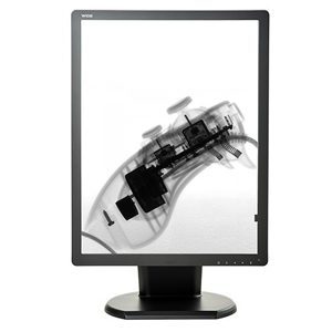
Requirements for Remote Radiology Monitors
Radiology professionals risk missing key information, and also risk providing an incorrect diagnosis, if they are evaluating images and scans without the correct DICOM calibrated display. This is something that has been studied in depth for diagnostic imaging within the enterprise, and remains especially important for a remote office location. According to a white paper on Teleradiology published by the American College of Radiology (ACR), radiologists must use one high set of standards “for both Teleradiology providers and on-site radiologists”, as doing anything less could lead to compromised patient care. These standards cover display luminance, pixel pitch, and display size, and also ensure DICOM calibration.
Luminance
Luminance is a measure of how much photon, or light energy, reaches the eye, and is measured in candelas per meter squared (cd/m²). A spectrum of precisely calibrated luminance levels including brightest white, lowest black, and all levels in between, ensures subtle abnormalities are visible and discernible. According to ACR standards , the minimum calibrated luminance is 350 cd/m² for radiology modalities including X-Ray, CT, MR, PET, US, and NM, and 420 cd/m² for Mammography and Tomo. In addition to ensuring your remote radiology monitor meets this minimum standard, it is also important to ensure the luminance output is stable over time. Medical displays have built-in backlight stabilization and monitoring. Our eyes don’t notice as a display slowly degrades over time or loses brightness. Backlight stabilization, a specification included in most radiology monitors, ensures the brightness level stays consistent over time, from monitor to monitor, and also controls the temperature at which the display runs.
Pixel pitch and display size
Individual pixels on a display determine the resolution of the monitor, with pixel pitch, or the density of pixels being a primary factor. Measured in millimeters from the center of one pixel to the center of an adjacent pixel, pixel pitch must be substantial in remote radiology monitors to provide the needed level of detail — approximately 200μm for most medical-grade displays.
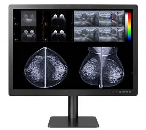
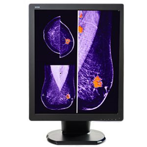
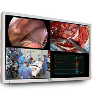
Another way to think about resolution is to consider the matrix size, or pixels per millimeter of screen space. The recommendation from the ACR for diagnostic reading is 2.5 line pairs/mm minimum, equivalent to a 3 megapixel (3MP) 12.3” diagonal display. For mammography/tomography, 5MP monitors, 8.8MP large format displays, or 12MP large format monitors are the standards that typically carry FDA clearance for breast imaging.
Display size is also important to reduce the need for zooming in and panning out repeatedly. An appropriate diagonal display size is 21” diagonal for 5MP and 30-32” for large-format 8.8MP or 12MP displays.
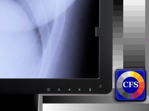
DICOM calibration
Radiology monitors used for at-home diagnostic imaging still need to meet the same standards they would inside Enterprise walls; they must be regularly calibrated to meet DICOM and ACR standards. Without meeting DICOM standards, diagnostic data is easily missed or misinterpreted – gray scaling and shadowing is truly lost. DICOM calibration ensures the diagnostic data output from the display is perceptually linearized, enabling the human eye to discern all gray levels. The user can be assured the diagnostic data is consistent on each display and from workstation to workstation throughout the enterprise.
Manual calibration with a handheld photometer and software combination is tedious and time consuming. Medical-grade displays used in the home office should ideally include built-in front sensors for luminance level calibration, rear backlight sensors to maintain stability continually and over time, and automatic calibration software. Calibration software should provide the ability to calibrate during convenient times (such as off-hours), send e-mail alerts for out-of-compliance issues, automatically diagnose and correct non-compliance issues if generate automated reports confirming adherence to the ACR, DICOM 3.14, and state specific requirements. A comprehensive, diagnostic quality hardware and software display combination is an invaluable tool for radiologists to feel confident in providing accurate diagnoses from a home office or remote location.
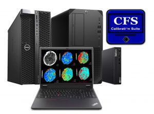
The DBI calibration software suite pairs your local display with a web based network administration option. Displays can be calibrated remotely and automatically to the DICOM grayscale display function and checked regularly for continued optimum performance. Automatic e-mail alerting for nonconformance and report generation are also included.
Setting Up the Right at Home Radiology Workstation
In addition to ensuring displays meet ACR standards and produce the least amount of eyestrain, a radiologist should make sure his or her remote radiology workstation is set up with proper ergonomics in mind. Repetitive strain injuries are especially common with radiology professionals due to the hours spent in one position while viewing studies.
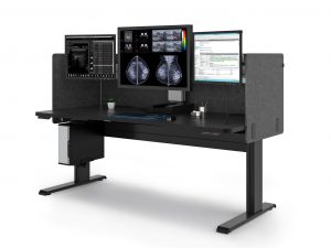
Countering the stress on the body from sitting in one place, engaging in repetitive motions to work a mouse and keyboard, or viewing monitors at an incorrect angle, is essential. The ergonomic radiology workstation should include options for sitting or standing, the ability to adjust the monitor height or keyboard position, and also allow for multiple display configurations.
Lighting should be carefully considered due to the clear effects on diagnostic image representation. Excess ambient lighting reduces image contrast, creating a “washed out” appearance, thus reducing your ability to discern subtle luminance level differences on even the best monitors. Screen-glare contributes to eye fatigue and strain. Controlling ambient room lighting while adding proper task lighting are key to delivering the highest quality image performance with the most comfortable viewing experience.
Ergonomic Radiology Workstation Requirements
Confidence and comfort are necessary for Radiologists to successfully read from home. Valuable time adjusting workstations coupled with image quality concerns are counterintuitive to efficiently reading remotely or from home. Double Black Imaging can assist in designing the best home workstation to meet your individual needs. With cutting edge display technology, automated calibration software, and on-call customer service and support, we ensure your home office is quickly up and running.
Have a home workstation and find your workspace is not optimized ergonomically? We are happy to speak with you and create a custom solution that fits your equipment needs, space requirements, and budget.
Double Black Imaging provides ergonomic radiology workstations and peripherals that ensure the right solution for your individual needs. With optimized ergonomics, your productivity will increase while eye strain and fatigue decrease.
Double Black Imaging has developed specially priced radiology workstation bundles to assist radiologists with home office needs during this COVID-19 pandemic. Learn more here!

 Radiology professionals today can do more than just look at X-Rays. They have a window into the human body with technologies like magnetic resonance imaging, positron emission tomography, and computed tomography, among many modalities. The technology that drives accurate and consistent medical imaging has grown and improved steadily, making it easier for radiology professionals to identify the correct diagnoses. Research dollars have been poured into methods for non-invasive, early discovery of a host of diseases, including cancer, stroke, and cardiovascular system conditions like artery blockages.