 Tax time will be here before you know it. Now is a good time to start pulling together your business expenses and documentation from this past year to ensure you are capitalizing on all the tax deductions you are entitled to as a business owner. The Section 179 tax deduction is an important tax saver for business owners that should not be overlooked.
This deduction can and does change from year to year (and has even changed within a tax year). Therefore, it is important to be aware of your deduction limit each year including the bonus depreciation which may also change depending on the tax year.
What is the Section 179 Tax Deduction?
The Section 179 tax deduction is a section of the IRS tax code originally designed to provide incentives and tax relief for small businesses although large businesses may also benefit from this deduction.
Section 179 “allows businesses to deduct the full purchase price of qualifying equipment and/or software purchased or financed during the tax year. That means that if you buy (or lease) a piece of qualifying equipment, you can deduct the FULL PURCHASE PRICE from your gross income.” This U.S. government incentive is designed to encourage businesses to buy the equipment they need while investing in themselves and their businesses.
Who Qualifies for the Section 179 Tax Deduction?
For the 2020 tax year, the good news is all businesses that finance, lease, or purchase used or new equipment outright totaling less than $3,630,000 should qualify for the Section 179 deduction.
More specifically, $1,040,000 of business assets qualify for the complete Section 179 deduction in 2020. That amount is reduced dollar for dollar when the amount of qualified assets used for business purposes reaches $2,590,000.
What Requirements Must Be Met to Claim the Section 179 Deduction?
Any equipment, vehicles, or software claimed as a tax deduction under Section 179 that is used for both personal and business purposes (such as cell phones for example) must meet the “More Than 50 Percent Business-Use” requirement. This means that the equipment, vehicles, or software being claimed must be used for business purposes more than 50% of the time that it is used for it to qualify for the Section 179 tax deduction.
What Equipment Qualifies for the Section 179 Deduction?
The following equipment “generally” qualifies for the Section 179 deduction and may be new or used—however it must be “new” to you as a business owner for it to be claimed as a deduction. It must also be purchased (and used) between January 1 and December 31 of the tax year in which you are wanting to claim it as a tax deduction against your business. The term “purchased” includes equipment you have leased, financed, or purchased outright with your own funds.
Equipment that generally qualifies includes:
- •Machines and a variety of equipment purchased for business use such as computers and “off-the-shelf” computer software.
- •Equipment purchased and used for business and personal use (the section 179 tax deduction is based on the percentage of time you use this equipment for business purposes).
- •Office equipment and furniture.
- •Different types of tangible personal property used for business purposes.
- •Specific types of improvements to existing non-residential buildings such as roofing improvements, fire suppression installations, security and alarm systems, and heating, ventilation, and air conditioning improvements.
- •Non-structural equipment and property attached to the building where you do business.
- •Vehicles used for business purposes (gross vehicle weight must exceed 6,000 lbs).
What’s the Difference Between Section 179 and Bonus Depreciation?
Bonus depreciation is taken after the Section 179 deduction is applied. Bonus depreciation usually benefits larger businesses that exceed the spending cap for new capital equipment in a given tax year (currently $2,590,000 for 2020).
It is also important to note that bonus depreciation is not offered every tax year. However, it is available for the 2020 tax year at a rate of 100%.
The most notable change for bonus depreciation in 2020 is that it may be applied to both new and used equipment, as long as the equipment is new to the business owner. This is a change from previous years in which bonus depreciation could only be applied to new equipment.
Businesses that experience a net loss this past year still qualify to deduct some of the cost of the equipment they purchased and then they may carry-forward the remaining loss to the following tax year. Similarly, if a business has no taxable profit during this current tax year, it may carry forward the loss experienced on equipment purchases to the subsequent tax year.
Is There a Way to Maximize My Tax Savings with Section 179?
Yes there is! One of the best ways to maximize your tax savings in 2020 is to lease or finance equipment purchases according to the Section 179 Qualified Financing rules. This is because as a business owner, you will only have to make the required lease or financing payments for the equipment during the year. However, you are entitled to deduct the full purchase price of the equipment you purchased in 2020, regardless of whether it is new or used.
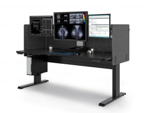
In effect, financing or leasing equipment and then taking the full Section 179 tax deduction can easily help business owners save more money in taxes than they spent in finance or lease payments throughout the year. Double Black Imaging offers a variety of leasing and financing solutions for our display systems, ergonomic workstations and CPU options!
Is It Possible to Preserve the Cash Flow in My Business and Still Take the Section 179 Deduction?
Absolutely! This has been and continues to be a year filled with uncertainty. Protecting cash flow for unanticipated expenses is a wise business idea. As discussed above, choosing to purchase equipment by leasing it can also put more tax dollars back in your business.
To keep lease payments as low as possible to preserve cash flow, consider asking about a non-tax capital lease. This type of lease will allow you to make smaller payments, protect your cash flow, and allow you to write-off the full price of the equipment you purchased up to the deduction limit— all while making the smallest payments possible.
How Do I Calculate My Potential Savings Using Section 179 on My Next Purchase?
That’s a great question. Fortunately, Section 179.org has an easy calculator that can help you estimate your potential savings if you are considering buying equipment this year.
To give you an idea of just how significant the tax and equipment savings can be for a business owner, Section 179.org provides this example:
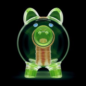
“Using a $75,000 equipment cost for a sample calculation shows how taking advantage of the Section 179 Deduction can significantly lower the true cost of the equipment purchased, financed or leased. In our example, $75,000 in equipment purchased has a true cost of $48,750. That’s $26,250 saved. Would you like an extra “25 grand-plus” this year on equipment you needed anyway?”
How Do I Ensure I Get the Section 179 Tax Deduction?
It is important to know you must elect to take the Section 179 tax deduction for business equipment you have purchased, leased, or financed during the calendar year. It is NOT automatically applied to your tax return.
To elect to take the Section 179 deduction, complete Part 1 of Form 4562 from the IRS and attach it to your tax return. Or speak with your tax preparer and tell them you want to elect to take the Section 179 deduction.
In addition, the Section 179 deduction is taken on an “item-by-item” basis. Therefore, you can choose to claim the deduction for only some of the equipment and/or software you purchased and not all of it if you choose.
Can You Use Section 179 Every Tax Year?
Yes, the Section 179 tax deduction can be used for business expenses and depreciation every year that you incur these expenses in your business. However, it is important to keep in mind that changes are made to Section 179 every year, and sometimes throughout the year. Therefore, be sure to stay informed about these tax code changes, what equipment, property, and improvements qualify, and what deductions you might be entitled to.
Be sure to speak to your tax preparer if you are not sure what deductions you are entitled to or prior to making large equipment or software purchases for your business to determine the most tax efficient way to do this.
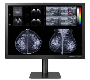
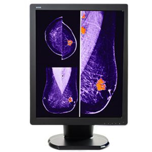
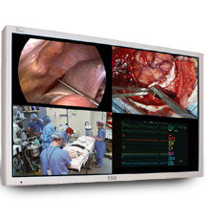
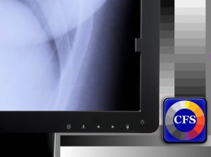
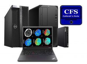
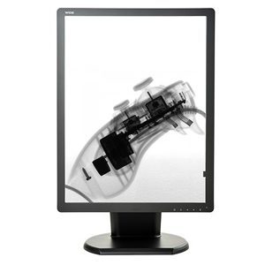
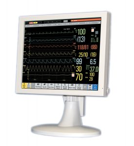
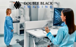
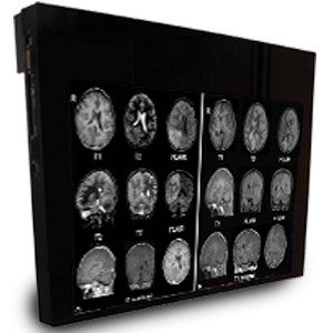
Double Black Imaging provides business owners with medical displays for PACS, monitors, and software as well as ergonomic workstations that qualify for the Section 179 deduction. Contact us at (844) 879-2247 with your business needs today and be sure to tell our diagnostic imaging specialists you want your purchase to qualify for the Section 179 deduction.

Source List:
https://www.section179.org
https://www.irs.gov/pub/irs-dft/i4562–dft.pdf
https://www.crestcapital.com/section_179_bonus?_ga=2.120781388.519583165.1608581164-847429404.1608581164
 Increase Patient Engagement: An interactive process with patients ensures a quality-based experience for them. Guiding them through their care journey at your healthcare facility while keeping an open communication will keep your patients engaged as well as deliver a personalized end-to end care.
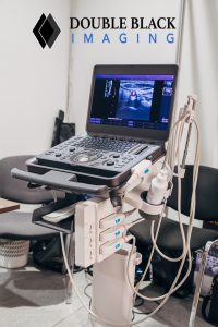
 Double Black Imaging offers the latest mobile medical imaging equipment at competitive prices and can walk you through how to implement it in your healthcare organization.
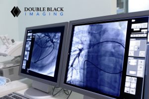
 Whether you’re looking to implement AI into your medical processes or furnish your healthcare facility, Double Black Imaging offers the latest technology at competitive prices and can help install your new device.

 The sedentary nature in radiology contributes to stress which has been shown to decrease job satisfaction and lead to poor mental health outcomes. However, it’s also thought that stress associated with sedentary radiology work may contribute to poor workstation ergonomics. This may lead to an array of work-related musculoskeletal injuries that 30%-60% of radiologists report such as eyestrain, neck & back pain, carpal tunnel syndrome, and headaches.
 Painter shares the following case of a neuroradiologist who faced a lawsuit related to work she performed at home: