
Why sRGB Calibration Is Necessary for Diagnostic Displays in Digital Pathology WSI
Executive Summary
Whole slide imaging (WSI) workflows rely on color‑accurate visualization for primary diagnosis, consultation, and research. Digital Pathology workflows depend on controlled staining, scanning, and image presentation, however variations can occur throughout the imaging chain. If the display does not present colors consistently and predictably, it introduces another source of variability and diagnostic reliability can be compromised.
sRGB calibration provides a universal, reproducible color baseline that aligns scanners, PACS/viewers, remote workstations, and cloud platforms. By keeping color rendering consistent across displays and locations, sRGB calibration reduces display-related variability, supports consistent image presentation and protects clinical workflows from unpredictable color drift.
The Digital Pathology Color Chain
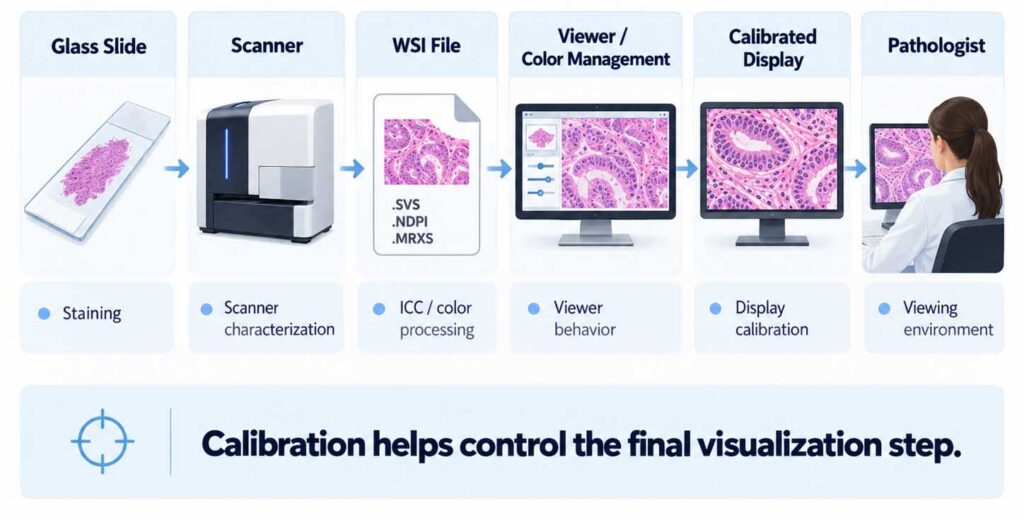
1. Introduction: The Critical Role of Color in Digital Pathology
Digital pathology imaging is uniquely sensitive to even small color differences. Pathologists rely on subtle visual cues such as:
- Hematoxylin and eosin saturation
- Nuclear detail visibility
- Cytoplasmic tone and contrast
- Detection of rare events or faint markers
- Identification of staining artifacts
These cues are highly dependent on stable, reproducible color rendering. Even minor shifts introduced by uncalibrated displays can alter perceived morphology and tissue interpretation.
Unlike radiology—where grayscale standards dominate—digital pathology is fundamentally a color‑driven diagnostic modality, making standardized color management essential.
Same WSI, Different Display Behavior
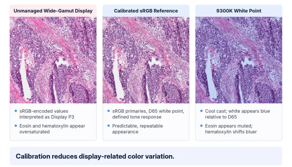
Simulated from one image tile. Left: an uncalibrated wide-gamut monitor with no color management. Right: a monitor set to a bluish 9300K white point instead of the D65 standard. Because you are viewing all three panels on your own screen or in print, the figure shows the type of each error, not its exact appearance. Image: TCGA-A6-2686-01A-01-01Z-00-DX1, The Cancer Genome Atlas, via NCI Imaging Data Commons, CC BY 3.0.
2. Industry Reality: sRGB as the Default Color Space
sRGB is widely supported across the digital pathology ecosystem as a reference color space for display workflows. In digital pathology, relevant components may include:
- Whole slide scanners (major vendors)
- Image management systems (IMS)
- Cloud pathology platforms
- PACS and universal viewers
- Research tools and machine‑learning pipelines
As WSI systems can use ICC profiles, embed color information, viewer-level color management, or proprietary platforms predictable display behavior is important. A display that is uncalibrated or deviates can produce inaccurate or unpredictable color reproduction.
Why sRGB?
sRGB was developed as a common reference color space for the web, photography, and general computing, and is standardized as IEC 61966-2-1:1999 (Amendment 1, 2003). Its strengths in pathology include:
- A defined tone response curve: a linear segment near black, then a 2.4 exponent, approximating gamma 2.2 overall.
- Predictable color primaries and a D65 white point
- Broad compatibility across imaging pipelines
- Stable, repeatable results independent of vendor
Calibrating diagnostic displays to sRGB removes the display as a source of inconstancy in the end-to-end pathology workflow.
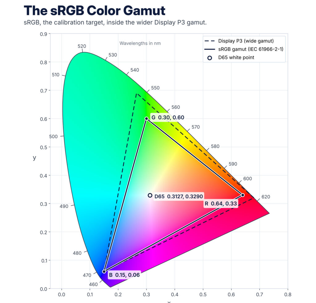
CIE 1931 xy chromaticity diagram. Spectral locus: CIE 1931 2o standard observer. sRGB primaries and D65 white point per IEC 61966-2-1:1999. Display P3 primaries: R 0.680, 0.320; G 0.265, 0.690; B: 0.150, 0.60. All colors in this diagram are approximate. The image is encoded in sRGB, so areas outside the sRGB triangle are shown as the nearest sRGB color. No display or printing process can reproduce every visible color, because any set of three primaries covers only part of the diagram.
3. Sources of Color Variability Without sRGB Calibration
3.1 Display to Display Variability
Off‑the‑shelf monitors—even high‑end panels—exhibit significant variation in:
- Native color gamut
- Gamma response
- White point (can vary from 5500K–8500K)
- Luminance uniformity
- Age‑related drift
Without calibration, two pathologists viewing the same slide on different displays may encounter differences, such as:
- Different hematoxylin intensities
- Incorrect eosin balance (too pink, too pale, or orange‑shifted)
- Over/under‑saturated chromogens in IHC
- Loss of fine contrast in nuclei
3.2 Scanner, Viewer and Color-Management Assumptions
WSI scanners and viewing applications may use embedded color information, ICC profiles or proprietary color transformations. Many profiles assume an sRGB display. When viewed on a wider‑gamut, uncalibrated, or inconsistently calibrated display:
- Color profiles are misinterpreted, ignored or misapplied
- Hues and saturations are amplified unintentionally
- Tissue tones appear unnatural or clinically misleading
3.3 Environmental and Aging Effects
Displays drift due to:
- LED aging
- Temperature fluctuations
- Backlight instability
Ongoing sRGB calibration and QA help detect and correct display drift, supporting long‑term consistency.
4. Clinical Impact of Non Standardized Color
4.1 Diagnostic Variability
Reviews and technical guidance show that color variability can affect the perceived appearance of diagnostically relevant features (Clarke and Treanor, 2017; Xiong and Sirintrapun, 2026), including:
- Grading dysplasia
- Breast cancer IHC scoring (ER/PR/HER2)
- Identifying mitotic figures
- Recognizing infectious organisms or rare events
Even relatively small shifts in color temperature, saturation, luminance or contrast can affect diagnostic confidence and consistency.
4.2 Remote Work and Telepathology
Remote consultations assume color parity across locations. A common, validated display target helps reduce disagreements caused by workstation factors rather than the underlying pathology.
4.3 Training, Research, and AI
AI models are highly sensitive to color variation in the image data itself. Display calibration has a different role: it supports consistency when human reviewers create annotations, labels, reference standards, or adjudications used in research and AI workflows. Non-standardized displays instead affect the reviewers and are likely to:
- Reduce reproducibility in digital pathology research
- Lead to inconsistent labeling during training
- Increase annotation noise
sRGB based display calibration can provide a stable viewing reference where sRGB is appropriate to the validated workflow.
5. Regulatory and Quality Considerations
Regulatory and professional guidance emphasizes validation, color reproducibility, and ongoing quality assurance for WSI systems and their viewing environments.
5.1 FDA Expectations (WSI Systems)
FDA‑cleared WSI systems are currently validated as complete imaging systems, with display-related performance considerations that include:
- Luminance
- Color response
- Gamma
- White point
While FDA 2016 guidance does not specifically name sRGB, it asks each manufacturer to define and justify its own color reproduction target. In practice, cleared pathology displays converge on sRGB. Filings for FDA cleared displays all list sRGB and a 6500 (D65) White Point. Calibrated luminance targets typically range from 370 cd/m2 to 500 cd/m2.
5.2 CAP/CLIA Quality Programs
CAP’s guideline update on WSI validation (Evans et al., 2022) requires laboratories to address:
- Validation of diagnostic equivalence between optical and digital workflows
- Changes to a validated system, such as replacing a display
A controlled, documented display environment (ie. sRGB calibration) supports these validation and QA activities by reducing avoidable display-related variability
6. Why Not Wide Gamut or DCI P3 for Pathology?
While wide‑gamut displays can reproduce more colors, they create major compatibility issues unless tightly managed.
Problems with wide‑gamut displays in pathology:
- Uncalibrated wide gamuts oversaturate H&E
- Small shifts produce exaggerated IHC chromogen intensity
- Color management becomes complex and brittle
- Vendors do not deliver WSI content in these gamuts
Unless the entire ecosystem shifts to a new standard—which would require scanner, viewer, ICC, regulatory, and workflow changes—sRGB remains the baseline.
7. Benefits of Calibrating Pathology Displays to sRGB
7.1 Diagnostic Consistency Across Workstations
Every calibrated workstation renders the same image values to the same color targets. Pathologists, regardless of location, are assured a visual baseline.
7.2 Interoperability Across Vendors
Scanners → IMS → PACS → display all behave predictably.
7.3 Reduced Training and Revalidation Burden
Fewer workflow differences to reconcile.
7.4 Accurate Reproduction of Scanner Intent
Display calibration helps reproduce the intended appearance of the scanner and viewer pipeline more consistently.
7.5 Long Term Stability Through QA
Displays with build in front sensors run QA on a schedule to detect any changes or drift and correct the display automatically.
8. Recommended Calibration Targets for Digital Pathology
A standardized calibration approach typically includes:
- Color Space: sRGB
- White Point: D65
- Gamma: 2.2 (sRGB TRC)
- Luminance: ~370–500 cd/m² based on display targets listed in FDA Clearances
- Black Level: As low as the display allows while preserving low-level detail
- Uniformity Compensation: Enabled where available
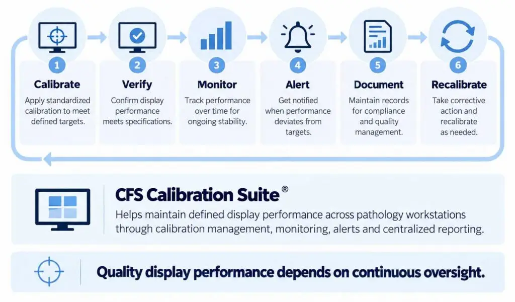
- Regular QA Intervals: Monthly or automated continuous monitoring
Calibration is not a one-time event
Regular QA maintains display performance over time.
9. Conclusion
Much of the digital pathology ecosystem is built around sRGB as the baseline for image creation, distribution, and display. Without sRGB calibration, diagnostic displays introduce unpredictable color shifts that undermine accuracy, consistency, and trust in WSI workflows.
Aligning displays to sRGB removes the display as a variable between what the scanner records and what the pathologist sees – supporting safe, reliable, and diagnostically sound digital pathology practice.
.
10. Authoritative References for sRGB Calibration & Color Standardization in Digital Pathology
Regulatory & Standards
- S. Food and Drug Administration (FDA).
Technical Performance Assessment of Digital Pathology Whole Slide Imaging Devices – Guidance for Industry and FDA Staff.
FDA, 2023 or April, 2016? - College of American Pathologists (CAP).
CAP Laboratory Accreditation Program Checklists – Digital Pathology Requirements.
CAP, current edition. - Digital Imaging and Communications in Medicine (DICOM).
DICOM Supplement 145: Whole Slide Microscopic Imaging.
NEMA, 2010.
- Evans A.J., et al. (College of American Pathologists).
Validating whole slide imaging systems for diagnostic purposes in pathology: guideline update from the College of American Pathologists in collaboration with the American Society for Clinical Pathology and the Association for Pathology Informatics.
Archives of Pathology & Laboratory Medicine, 2022;146(4):440-450. - U.S. Food and Drug Administration (FDA).
510(k) decision summaries for digital pathology displays
Peer-Reviewed Scientific Literature
- 4. Yagi Y.
Color standardization and optimization in whole slide imaging.
Diagnostic Pathology, 6(S1): S15. 2011. - García-Rojo M.
International clinical guidelines for the validation of digital pathology systems for primary diagnosis.
Journal of Pathology Informatics, 2016;7:17. - Pantanowitz L., et al.
Whole Slide Imaging for Primary Diagnosis: A Systematic Review.
Archives of Pathology & Laboratory Medicine, 2020;144(6): 672-688. - Mukhopadhyay S., et al.
Whole slide imaging versus microscopy: Systematic review and meta-analysis of diagnostic equivalence.
Human Pathology, 2018;72: 1–11. - Baidoshvili A., et al.
Evaluating the influence of color calibration in digital pathology.
Journal of Pathology Informatics, 2013;4:15. - Rojo M.G., Bueno G., Slodkowska J.
Review of imaging solutions in digital pathology.
Journal of Biomedical Informatics, 2008;41(4): 552–562. - Clarke E.L., Treanor D.
Colour in digital pathology: a review.
Histopathology, 2017;70(2):153-16 - Xiong Y., Sirintrapun S.J.
Display performance and standards for primary digital pathology sign-out: technical specifications, validation, and quality assurance.
Journal of Pathology Informatics, 2026. PMCID: PMC13262270
Color Science & Imaging References
- IEC 61966-2-1:1999.
Multimedia systems and equipment — Colour measurement and management — Part 2-1: Colour management — Default RGB colour space (sRGB). - Stone J., et al.
Device color profiles and the importance of sRGB consistency in medical imaging.
Journal of Imaging Science and Technology, 2013. - Bernas T., et al.
Color and intensity variation in microscopy: sources and solutions.
Cytometry Part A, 2014;85(3): 231–241.
Authored by: Dave Lemke, Double Black Imaging