Category: Radiology & Diagnostics

15 Jun 2024

Чому варто обрати Slotoking?

Casino

Slotoking являє собою світ азарту та веселощів, де кожен гравець може знайти щось за своїм смаком. Це не просто казино — це дійсно унікальний досвід, який залишає незабутні враження. З першого ж кроку на платформу, ви поринете у всесвіт азартних ігор, де все створено для вашого комфорту та задоволення.

Переваги Slotoking

Є безліч причин, чому Slotoking виділяється серед інших казино, ось деякі з них:

  • Великий асортимент ігор, який постійно поповнюється.
  • Зручний та інтуїтивно зрозумілий інтерфейс користувача.
  • Швидкі та безпечні транзакції.
  • Професійна служба підтримки 24/7.
  • Щедрі бонуси та акції для нових і постійних гравців.

Кожна з цих особливостей робить Slotoking ідеальним вибором для тих, хто цінує якість і зручність. Ваша гра не буде обтяжена незрозумілим управлінням або проблемами з виведенням коштів. Натискаючи кілька кнопок, ви швидко знайдете свою улюблену гру, отримаєте бонуси та зможете спілкуватися з підтримкою в разі необхідності.

Ігрові можливості

Асортимент ігор у Slotoking вражає своєю різноманітністю. Чи ви є шанувальником класичних слотів, чи надаєте перевагу настільним іграм, таким як покер чи рулетка, Slotoking має все, що потрібно для чудового дозвілля. Кількість ігор постійно зростає, що означає, що ви ніколи не відчуєте нудьги, адже розробники програмного забезпечення постійно працюють над новими та захопливими проєктами.

Slotoking співпрацює лише з найкращими виробниками ігор, такі як NetEnt, Microgaming, Playtech, які забезпечують якісний та чесний геймплей. Всі ігрові автомати мають ліцензії, що гарантує чесність ігор. Можливості азартних розваг включають не тільки слоти, але й живе казино, де ви зможете відчути справжню атмосферу гри з реальними дилерами.

Безпека та надійність

Slotoking активно піклується про безпеку своїх користувачів, застосовуючи найсучасніші технології для захисту даних та фінансових операцій. Ваша інформація завжди буде в безпеці, адже казино працює лише з перевіреними платіжними системами, що гарантує безпеку кожної транзакції. Не менш важливою є конфіденційність даних гравців — ви можете бути впевнені, що ваша особиста інформація не буде передана третім особам.

Процес верифікації даних гравців здійснюється з використанням найновіших технологій захисту, щоб переконатись у дотриманні всіх законодавчих норм. Slotoking також забезпечує відповідальне ставлення до гри, пропонуючи інструменти для контролю над власними витратами та безпеки.

Бонуси та акції

Slotoking завжди радує своїх клієнтів різноманітними бонусами. Будь то вітальні бонуси для новачків чи спеціальні пропозиції для постійних гравців, ви завжди знайдете щось вигідне. Ми пропонуємо не лише одноразові бонуси, але й програми лояльності, де кожен рівень приносить додаткові переваги та спеціальні пропозиції для найактивніших гравців. Розгляньте таблицю нижче, щоб дізнатися більше про актуальні акції.

Бонус Умови отримання Період дії
Вітальний бонус Реєстрація та перший депозит Постійний
Бонус на депозит Поповнення рахунку До кінця місяця
Кешбек Повернення частини програної суми Щотижня
Різдвяний бонус Участь у акції різдвяних тижнів Грудень 2023
Літній марафон Гра у вибрані слоти протягом літа Червень-Август

Кожен бонус — це унікальна можливість спробувати різні види ігор або ж додатково збільшити свій виграш. Завітайте на https://aska.com.ua/ та дізнайтеся більше про можливості, які відкриває перед вами Slotoking. Ми впевнені, що ви залишитеся задоволені своїм вибором і насолодитеся комфортним грою в унікальній атмосфері Slotoking.

Отже, не втрачайте шанс стати частиною світу яскравих емоцій та адреналіну з Slotoking. Завантажте мобільний додаток або грайте онлайн – ваше задоволення вже чекає. Завдяки сучасним технологіям, гри в Slotoking доступні з будь-якого пристрою — чи то мобільний телефон, планшет, чи комп’ютер. Графіка та анімації ігор адаптовані для будь-якого екрану, забезпечуючи максимально приємний досвід.

Приєднуйтесь до спільноти гравців, які вже відкрили для себе всі переваги Slotoking, і отримайте незабутні враження від гри. Будьте частиною наших святкових заходів, турнірів та лотерей, які роблять вашу гру ще більш захопливою. Заходьте, грайте, вигравайте — і пам’ятайте, що Slotoking — це місце, де ваші мрії можуть стати реальністю!

17 May 2024

Неймовірний Досвід у Pokermatch

Гра в казино завжди була настільки ж захоплюючою, як і захоплюючою. І коли мова йде про Pokermatch, ми говоримо про місце, що перевершує всі очікування. Pokermatch пропонує все, на що може сподіватися любитель азарту. Від неперевершених ігор до розкішної обстановки – це казино має все.

Casino Experience

Одним з ключових факторів, які роблять Pokermatch особливим, є його неймовірний вибір ігор. Тут ви знайдете:

  • Класичні настільні ігри: покер, блекджек, рулетка;
  • Сучасні слоти з різноманітними тематиками та бонусами;
  • Живе казино з реальними круп’є;
  • Ексклюзивні турніри та змагання;
  • Прогресивні джекпоти, що ростуть щодня.

Крім великого вибору ігор, Pokermatch також відоме своїм відмінним обслуговуванням клієнтів. Команда підтримки завжди готова допомогти з будь-якими питаннями чи проблемами, які можуть виникнути у гравців. Більше того, Pokermatch працює цілодобово, надаючи кожному клієнту можливість насолоджуватися іграми в зручний для нього час.

І тут все не лише про ігри. Обов’язково варто згадати про розкішні ресторани та бари, які пропонують широкий вибір страв та напоїв. Атмосфера тут завжди чудова, а введені COVID-19 заходи безпеки гарантують безпечний відпочинок для всіх відвідувачів.

Що робить Pokermatch ще більш привабливим, це його відданість забезпеченню чудових бонусів та акцій. Кожен новий клієнт отримує шанс скористатися ексклюзивними бонусами, які можуть значно збільшити банкрол. Регулярні акції для постійних гравців також роблять гру ще більш вигідною.

Переваги гри в Pokermatch

Перевага Опис
Великий вибір ігор Від класичних настільних ігор до новітніх слотів та джекпотів
Безпека та захист Найкращі технології шифрування даних для вашої безпеки
Цілодобова підтримка Дружня клієнтська підтримка готова допомогти в будь-який час
Бонуси та акції Щедрі бонуси для нових клієнтів та регулярні пропозиції для постійних гравців

Pokermatch розташований у самому центрі міста, забезпечуючи легкий доступ для всіх відвідувачів. Інфраструктура навколо казино розвинута та зручна для гостей, що приїжджають з інших міст чи країн. В комфортних номерах поруч із казино можна відпочити після насиченого дня гри, що ідеально підходить для тих, хто планує провести кілька днів у цьому дивовижному місці.

Важливість такого виду розважальних послуг важко переоцінити, адже казино давно стало одним із найпопулярніших способів проведення дозвілля. Завдяки сучасним технологіям, Pokermatch змогло об’єднати в собі традиційний підхід до гри з новітніми інноваціями, що робить досвід унікальним для кожного гравця. До прикладу, система винагород, впроваджена в казино, дозволяє накопичувати бали за гру, які можна обменяти на додаткові можливості або привілеї у різних зонах казино.

Окрім настільних ігор, слоти також користуються неабиякою популярністю серед відвідувачів. І це не дивно, адже розробники Pokermatch постійно працюють над оновленням існуючих ігор, а також додають новинки, що враховують сучасні тенденції індустрії. Користувачі можуть насолоджуватися не тільки традиційними слотами, а й вибором, що включає різноманітні тематичні автомати з кінематографічною графікою та захоплюючими сюжетами.

Особливу увагу варто приділити живим іграм, які стали доступні завдяки впровадженню новітніх технологій. Гравці можуть обрати стіл з живим дилером та насолоджуватися грою в режимі реального часу, що дарує неперевершене відчуття присутності у справжньому казино, навіть перебуваючи вдома. Якість трансляції на висоті, а професійні круп’є забезпечують гравцям найкращий сервіс.

Безумовно, не всі переваги гри в Pokermatch можна описати словами. Це місце потрібно відвідати особисто, щоб відчути всю енергію та азарт, що витає у повітрі, коли ви заходите до ігрової зали. І зрештою, натиснувши на https://chil.com.ua/, ви отримаєте можливість самостійно переконатися у всіх перевагах Pokermatch. Незалежно від того, чи шукаєте ви захоплюючі ігри, відмінне обслуговування або неперевершену атмосферу, це казино – ваш ідеальний вибір. Безпека, якість і задоволення – ось що гарантує Pokermatch для кожного свого клієнта.

Переходьте на сайт і відкрийте для себе світ гострих відчуттів та величезних можливостей разом із Pokermatch. Відчуйте емоції, адреналін та безліч позитивних вражень, які залишаться з вами надовго. Гарантія якості та задоволення від гри – це те, що робить відвідування Pokermatch справжнім святом.

11 May 2024

Дослідження світу розваг з Bet2fun

Casino Winning Banner
Радість перемоги з Bet2fun

Світ азартних ігор завжди захоплював уяву людьми, і з розвитком сучасних технологій, цей сектор зазнав значних змін. Однак, навіть у цифровий вік, існують заклади, які зберігають чарівність класичних казино, надаючи при цьому інноваційні рішення для гравців. І одним з таких виняткових закладів є Bet2fun. Відвідайте їх офіційний сайт за допомогою https://inpc.com.ua/ та переконайтесь самі.

Bet2fun – це місце, де зустрічаються азарт та грація. Від моменту входу віртуальні двері цього закладу відкривають перед гравцем нескінченні можливості. Наші головні переваги:

  • Вражаючий вибір ігор
  • Сучасні технології забезпечення безпеки
  • Приємні бонуси та акції

Гра в Bet2fun – це завжди захоплюючий досвід, завдяки сучасному підходу до розробки та постійного вдосконалення інтерфейсу. Ми прагнемо забезпечити найкраще обслуговування, щоб кожен момент проведений на нашій платформі був незабутнім. Крім того, ми завжди слідкуємо за останніми тенденціями в ігровій індустрії, щоб пропонувати вам найактуальніші новинки та інновації.

Ігровий асортимент: що пропонує Bet2fun?

Від класичних ігор до новітніх слотів, Bet2fun пишається своїм різноманітним асортиментом, що здатний задовольнити навіть найвибагливішого гравця. Наші гравці можуть насолодитися не лише традиційними азартними розвагами, але й ексклюзивними іграми, які ви не знайдете ніде в інших онлайн-казино. У нас доступні наступні категорії ігор:

Категорія Найпопулярніші ігри
Слоти Starburst, Mega Moolah
Карточні ігри Блекджек, Баккара
Рулетка Європейська рулетка, Французька рулетка

Високоякісна графіка та захоплюючий звук створюють незабутню атмосферу, що дозволяє відчути справжнє занурення в ігровий процес. Кожна гра проходить сувору перевірку якості та відповідності високим стандартам галузі, забезпечуючи неперевершений досвід для наших клієнтів.

Основна увага на безпеку та чесність

Bet2fun прагне підтримувати найвищі стандарти безпеки. Завдяки використанню передових технологій шифрування, всі клієнти можуть бути впевнені у повній конфіденційності своїх даних та фінансових операцій. Ми співпрацюємо з провідними постачальниками платіжних рішень, щоб гарантувати надійність та швидкість усіх транзакцій.

Крім того, казино пропонує прозорі механізми гри та співпрацює лише з ліцензованими постачальниками програмного забезпечення. Важливо, що всі ігри базуються на системах генератора випадкових чисел, що виключає можливість маніпуляцій результатами.

Також для нас важливо, щоб усі гравці мали можливість контролювати свій ігровий бюджет, тому ми впровадили спеціальні інструменти для відповідальної гри. Наші консультанти з відповідальної гри завжди готові надати допомогу та поради для підтримки здорового підходу до азартних ігор.

Вітальні бонуси та захоплюючі акції

Щоб зробити старт для новачків максимально приємним, ми підготували щедрі вітальні бонуси. Нові гравці можуть скористатися вступними пакетами, що включають безкоштовні обертання та бонусні кошти. Постійні гравці також не залишаються поза увагою завдяки регулярним акціям та спеціальним пропозиціям. Наші бонусні програми розроблені таким чином, щоб надати максимальну вигоду саме вам.

Навчальні матеріали та огляди ігор доступні на нашому сайті, щоб ви могли завжди залишатися обізнаними у світі азартних розваг. Вибір зроблений – приєднуйтесь до сім’ї Bet2fun вже сьогодні!

Чому обрати саме нас?

Наша команда постійно працює над покращенням сервісу та розширенням можливостей для наших клієнтів. Ми регулярно вдосконалюємо наш функціонал, щоб забезпечити гладкий і приємний геймплей, без затримок та технічних збоїв. Тому обравши Bet2fun, ви можете розраховувати на:

  • Високий рівень обслуговування
  • Миттєві виплати
  • Прогресивні джекпоти
  • Унікальні турніри та змагання

На завершення, наш сайт з чекає на вас – залишилося лише зробити перший крок! Нашіж консультанти підтримки завжди готові допомогти при будь-яких питаннях або труднощах, щоб забезпечити вам бездоганний досвід гри. Приєднуйтеся до нас, і ви не пошкодуєте про свій вибір! Відчуйте адреналін, виграші та справжнє задоволення разом з Bet2fun.

12 Sep 2022

New Advancements in X-Ray Imaging

Scintillators, nanophotonic scintillators, nanophotonics, and HiP-CT are just a few of the new terms you will hear when discussing new developments in X-ray imaging.

All of these new advances in radiology help medical professionals see with greater detail and to perform analyses within the body, within the organ of interest.

Scintillators have been in use for approximately 70 years and have been used to develop brighter and faster light emissions.  Now, scientists are manipulating length scale and changing the optical properties of the technology.

Nanophotonic scintillators are created by making patterns inside the scintillators or gluing another material with holes in it onto the nanoscale.  Researchers have been able to calculate the scintillation levels that would be produced by any configuration of nanophotonic structures.

The process is complicated, and the framework could integrate three different types of physics.  The effort has been worth it though, because Charles Roques-Carmes1 at MIT has reported that they “have found a good match between their predictions and the results of their subsequent experiments.”

The belief is that this will open a new field of research in nanophotonics – using research that has already been done in the field of nanophotonics to improve existing materials that scintillate.

Soljacic1 reported a tenfold improvement in emission but stated that it is possible to get up to 100 times improvement – and it is speculated that this will only lead to further improvement.

This is just one development that led to the European Synchrotron Radiation Facility (ESRF)’s Extremely Brilliant Source (EBS)2.  The goal of this research was to image intact human organs to the cellular level in three dimensions.  Their research enabled them to perform non-destructive, three-dimensional (3D) scans in whole human organs at any location.  This technology has been used to image five intact human organ types:  brain, lung, heart, kidney, and spleen.

Previous attempts to scan whole organs at the cellular level have shown promise.  However, they often require the use of stain or contrast material, take a long time to obtain the scan, and/or cause damage to the human tissue.

Synchrotron X-ray tomography (sCT) has been used to develop the EBS that achieves high resolution compared to the size of the biological sample.


What These X-Ray Advancements Mean For The Medical World


The researchers indicated that they believe “the improvements in medical diagnostic X-rays and CT scans, will reduce dose exposure, and improve image quality.”  This in turn will justify the extra time and effort required to integrate these scintillators into existing X-ray machines. These new scintillators could also enable faster, more accurate inspections.

It is speculated that the combination of nanophotonic and scintillators could help you achieve higher resolution, reduced X-ray dose and perform energy-resolved X-ray imaging.

This technique could facilitate understanding of system-level behaviors in health or disease as well.

Claire Walsh2, a mechanical engineer from University College London who is working on the project, speculates that linking HiP-CT images to clinical images through AI techniques will enable validation of ambiguous findings in clinical images with a high degree of accuracy.

Walsh also conjectures that the detailed imagery obtained from HiP-CT will be used with machine learning to improve information deduced from clinical imaging such as MRI and CT scans.  It will also be able to better calibrate and improve current technologies in use.


How It Can Help Deliver Better Patient Care


The most notable benefit of these advances in radiology is that the patient receives a more accurate diagnosis.

Additionally, the studies are performed at a higher speed with a lower dose of radiation.


This New HiP-CT Technique Came After The Beginning Of The Global Pandemic


The use of HiP-CT was significantly impacted by COVID-19 – in a positive way.  HiP-CT was used during the pandemic to provide new insights into how the disease disrupts blood oxygenation.

After the beginning of the pandemic, researchers began to use several techniques that were used at ESRF to image large fossils.   This was combined with the EBS to allow researchers to see the extremely small vessels within a complete human organ so that it could be distinguished from the surrounding tissue.  This 3D visualization even allowed them to observe specific cells.  The HiP-CT bridges the scales between CT and MRI scans which can resolve down to just below a millimeter and histology, electron microscopy and other similar techniques that resolve structures with sub-micron accuracy but require small biopsies of tissue from an organ.

It was known during COVID-19 that a fundamental pathological sign of the disease was a sharp drop in blood oxygenation levels.  HiP-CT provided the first direct evidence  that this is a result of “shunting” in the lungs.

Danny Jonigk2 at Hannover Medical School, a researcher working in the project, stated that they combined these molecular methods with the HiP-CT multiscale imaging in lungs affected by COVID-19 pneumonia and gained new insight as to how “shunting” occurs in COVID-19 injured lungs and the impact it has on oxygen levels and the circulatory system.

Researchers compared findings or orthogonal 2D slices through a COVID-19 affected lung with HiP-CT images of the same lung prior to the microscopic analysis.  They both identified cavitation of lung parenchyma, alveolar obstruction, thickening of septa between adjacent alveoli and blood capillary occlusion with adjacent cellular infiltrates.  A 3D quantitative analysis revealed a decrease in the surface area-to-volume ratio and increased septal thickness between control versus COVID patients.

This information was used to develop a fourth-generation synchrotron source which has enabled hierarchical 3D imaging of multiple intact human organs. The images generated have been high quality from whole organs down to individual organotypic functional units and certain specialized cells at any location within the organ.

The information gained from this research was used to develop The Human Atlas and Human Biomolecular Atlas which is an effort to map the hierarchical structure of human organs.


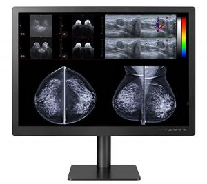
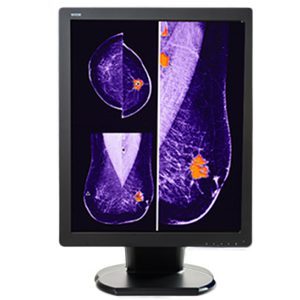
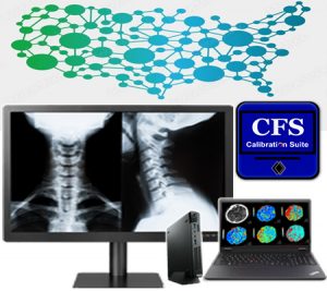
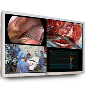
About Double Black Imaging

Double Black Imaging is based in the USA and creates 100% of our software and performs 100% of our display integration in the USA.  We are proud to be the largest medical display supplier and calibration software developer.

You can buy from us knowing that we have a reputation for the industry’s finest customer service, with acknowledgements from thousands of Radiologists and IT professionals.

We provide you with diagnostic imaging systems that keep you on the leading-edge of X-ray services while helping you be more efficient and contain healthcare costs.  We don’t just say these things, we mean them, and we stand by them.

Are you looking to add new X-ray services to your facility?  Contact us here, call us at (877) 852-2870, or email us at sales@doubleblackimaging.com to see how we can help you provide the best service to your patients.


Charles Roques-Carmes, Nicholas Rivera, Ali Ghorashi, Steven E. Kooi, Yi Yang, Zin Lin, Justin Beroz, Aviram Massuda, Jamison Sloan, Nicolas Romeo, Yang Yu, John D. Joannopoulos, Ido Kaminer, Steven G. Johnson, Marin Soljačić. A framework for scintillation in nanophotonics. Science, 2022; 375 (6583) DOI: 10.1126/science.abm9293


Walsh, C.L., Tafforeau, P., Wagner, W.L. et al. Imaging intact human organs with local resolution of cellular structures using hierarchical phase-contrast tomography. Nat Methods 18, 1532–1541 (2021). https://doi.org/10.1038/s41592-021-01317-x

18 Jul 2022

How Can Point of Care Technology Help with Your Radiology Department?

Point of Care Technology (POCT or POC), also referred to as Point of Care Testing, has significantly impacted medical care over the past 40 years.  We have seen care move from the hospital to the local clinic or laboratory to the patient’s home.

We have seen the tester change from doctor to nurse or technician, to patient, to machine.

The primary impact has been

  • To make receiving care faster for patients
  • To make care easier to use
  • To make testing and intervention more accurate
  • To provide greater satisfaction for patients receiving that intervention


What is Point of Care Technology?


“What is POC Technology?”  Put simply, POC stands for Point-of-Care.  This refers to whether the patient care is performed in a hospital, a clinic, a doctor’s office, a laboratory, or the patient’s home.

The goal of POC technology has been to provide the testing/treatment close to the patient’s home.

Two primary categories of POC Technologies are hand-held devices and bench-top devices.

Hand-held devices are now often come in the form of equipment that can attach to a phone or tablet and analyze samples (usually blood, urine, or saliva).

Bench-top devices are being miniaturized so that they take less space in the lab or office and can be brought to the patient.

Point of Care Technology has impacted every aspect of patient care, every specialization, and every location where services are provided.

As previously noted, this has made things easier for patients and caregivers, increased patient satisfaction and increased revenue across the board.


How Point-of-Care Technology Is Changing Radiology Departments


There continue to be some challenges that radiologists and clinicians encounter when implementing POC technologies in patient care. These are opportunities to expand service options and improve your current services and department functionality.

Implementation of portable radiology services in the hospital setting provides you opportunities to realize improvements in quality of care, patient satisfaction and hospital revenue.

It can seem overwhelming at first, even impossible. However, facilities that have embraced POCT report the benefits suggested above in this article.

Three ways POC imaging can positively impact your radiology department can be summarized as follows:

  • You can perform an increased number of studies
  • You will see improved surgical outcomes in the OR
  • You can have reduced exposure to risk factors encountered during patient transport

Let’s discuss these benefits further.


Impacting The Number Of Studies Performed


Portable imaging provides an opportunity to complete studies that were previously too complicated or risky.  With POC tests, you can bring the imaging to the patient and provide successful intervention to patients who previously could not come to the Radiology Department for care.

Further, you can include valuable patient data in your clinical research because you now have access to a population that previously could not be sampled.

Without a requirement for transportation, the procedure can be performed quickly and more efficiently.  Therefore, you can complete more procedures in the same amount of time and increase the number of procedures that can be completed because you can now treat patients who couldn’t travel to access your intervention.

Hospitals using POC imaging report an increased number of studies in several areas.  Among those are brain viability, intraoperative imaging, lymphoscintigraphy, GI Bleed, Lung perfusion, HIDA, MUGA and Bone Flow/Blood Pool.


Improving Surgical Outcomes In The OR


POC imaging is influencing surgery in the OR and improving outcomes.  This leads to improved quality of care and prevents readmission.

Two primary factors that impact surgical outcomes are the time under anesthesia and real-time results.

Increased time under anesthesia is known to increase the risk of surgical complications.  It follows then that decreasing the time under anesthesia can reduce that risk.

Portable imaging technology allows patients to be anesthetized for completing both imaging procedures and surgery in the OR.  Since patients don’t need to be anesthetized in while being transported from radiology to the surgery room, their hospitalization can be much safer because this risk has been minimized.

Patients can be more secure in the outcome of their procedure and feel greater satisfaction with it because the portable imaging device allows full confirmation of surgical success/accuracy during the procedure.  They can rest well knowing that the lesion or tumor removal was complete and that any remaining or unknown masses have been identified.


Reducing The Risks Of Patient Transportation Protocol


Many are surprised to learn of the number of risk factors encountered during patient transport.

Risks a patient can encounter during transport fall into several categories:

  • Exposure
  • Medical complications
  • Injury
  • Interruption of care/services

Patients can be exposed to infectious pathogens during transport or can experience variables that increase the risk of ventilator-acquired pneumonia.

Medical complications can range from hypoxemia, to tachycardia, to pulmonary complications, to arrhythmia, to cardiac arrest, just to name a few.

Injury, as well as interruption of care/services, can occur if equipment is dislodged, or patients are removed from services for the purpose of transport.

While these risks could occur at any stage during a hospitalization, the risk of complications is even greater if they occur during transport when the patient is away from the resources of his medical unit.

The risk is further increased if the patient is critically ill.  Difficult decisions might have to be made to forgo a needed medical procedure because the risks of transport are too high.

Transport of critically ill patients comes at a high cost when you consider that they often require a team of professionals and a variety of medical interventions (oxygen, suction, portable ventilation).

The high cost of transport is related to

  • Labor of this team of professionals
  • Risks to patient safety
  • Diverting care providers from the medical unit

Portable imaging can reduce these concerns by providing the services on the unit where transport is avoided and the only person needed is the medical technician performing the procedure.


Future Trends In POC Testing


It is clear from looking at the changes over the years and the benefits to patients, caregivers, and facilities, that POC testing will continue to make significant contributions to patient care, quality of services and your revenue.

You can easily see that new POC testing will continue to emerge as the needs, potential benefits, and means to achieve that POCT are identified.

Some areas where you can expect new POC Technologies to emerge are:  Virtual Reality and 3D imaging, Artificial Intelligence, Nuclear Imaging, Intraoperative imaging, Wearable Technology.


Work With Double Black Imaging For Your Medical Displays & Monitor Needs


Double Black Imaging works with you, the professionals who use our monitors, to develop state-of-the-art equipment that gives you portability when you need it.  We know portability is important.  We also know that portable equipment needs to retain the functionality demonstrated by the stationary equipment you previously have used to serve your patients.

We recognize the importance of trends in medical care and POC Technologies is just one of those trends that guides the development of our monitors.

Are you looking to improve your POC Testing and other technologies?  Let us help you configure a system that will meet your needs and your budget.  Contact us today at (720) 826-0859 or email us at sales@doubleblackimaging.com.

15 Jun 2022

What is DICOM in Medical Imaging?

DICOM stands for Digital Imaging and Communications in Medicine. It is an internationally standardized protocol for managing and transmitting images with related data.  It ensures interoperability of systems that produce, display, query, retrieve, share, send, store, print, and process medical images.


Why Is DICOM in Radiology Important?


Advances in imaging technology and increased use of computing in clinical work created a need for a standard method of transferring images and their associated information.  DICOM was developed to answer this need.  It can ensure this transfer between devices, regardless of vendor or manufacturer.

DICOM in radiology is important to maintain this accuracy because facilities rely on medical imaging for accurate communication and profit.

With DICOM it is easier for physicians to access images and reports, allowing access from in house or remote locations.  This greatly improves the efficiency, and therefore the quality, of patient care.

What Does A DICOM File Contain?


DICOM images consist of two components: a header and the image.

The header contains the data that describes the image, primarily patient data.  As with other medical files, this includes the patient’s demographic information such as their name, age, gender, and date of birth.

The header can also contain information about the image, such as acquisition parameters, pixel intensity, matrix size and dimensions of the image.


How To View A DICOM File


It is only possible to view DICOM images using special software mean specifically designed for viewing these files.  The software can be proprietary or third party.

Proprietary software is what is installed in the equipment you purchase for medical imaging.

You can view the images at the same workstation where the images, such as CT or MRI, were acquired by the machine using the installed DICOM viewer. It will allow you to view images sequentially and to reconstruct the images.

Unfortunately, the proprietary software only allows you to view the images in the same location as the hardware.  The only way you can transfer the images is to a portable storage device or a network; however, you need to compress the images to do that.  Compression and exporting images usually prohibits access to the original image.

Because of these and other issues, third-party software has been developed and is used more commonly.  Third-party applications allow you to open DICOM files from any source (PACS server, the internet, a CD, or a DVD).

There are many viewing applications available for DICOM in radiology. Each viewer has different features, and you can consider the features available in each application as they compare to your viewing needs. The availability of third-party applications facilitates interoperability of your medical viewing device with other devices.


What Can I Do To A DICOM File In Addition To Simply Viewing The Image?


DICOM medical image viewers provide functionality that can increase the efficiency and effectiveness of your work.  Some of the capabilities go beyond what was first imagined when imaging technology was developed.  Among the things you can achieve with DICOM are:

  • Comparison of medical images – With DICOM, you can view two digital images side by side to make comparisons.


  • Enhancement of image quality – DICOM allows you to zoom in to see detail and to change the contrast in an image. By increasing or decreasing the brightness of the image, you can make better distinctions between radiodensity and radiolucency in different areas of the image.


  • Reconstruction of images – This allows you to see aspects of the anatomy that are not available from the original images.
    • Two-dimensional to three-dimensional rendering is a method that takes the initial two-dimensional images from the initial DICOM data set that are taken in all three planes (axial, coronal, and sagittal) and reconstructs them into a three-dimensional rendering.
    • Multiplanar Reconstruction (MPR) uses the 3D reconstructed images and makes slices of them to allow you to view different anatomical levels at different angles than those acquired in the initial images.


  • Combination of images – Some DICOM applications allow you to combine different modalities, such as PET and CT images. Using this combined image, you can simultaneously leverage the advantages of both types of imaging modalities.  So, for example, a PET scan would locate areas of high metabolic activity, and you could combine that with the CT scan to map it to corresponding anatomical sites.


  • Measurement of image targets– If you want to measure the linear size or volume of anatomic structures, you will appreciate apps that have this functionality. Being able to make these measurements accurately and efficiently is useful in assessing treatment efficacy or in planning treatment.


Can You Transfer A DICOM File Into Other Formats?


Each DICOM file contains several high-resolution images.  Because this can create a very large file, compression is needed so you can transfer it.

It is important to note that compression of DICOM files uses two strategies:  lossless and lossy.

Lossless does not lose any information when it is compressed.  You can easily recover the original file at any time.  However, this requires a lot of processing and results slow file opening and saving.  A lot of compression is not possible with this strategy.

Lossy compression achieves greater compression because it removes some data.  Typically, redundant data is removed.  However, if a significant amount of compression is done, image quality can be impacted.

DICOM files can be compressed and exported into various formats, including JPEG, TIFF, PNG and GIF. This table summarizes the formats.


Other Capabilities of DICOM in Radiology


In addition to capabilities that enhance your viewing of and working with images, DICOM also has several functionalities that enhance the operation of your hospital or clinic.

DICOM supports network image management, network image interpretation management, network print management, image procedure management and offline storage media management.

DICOM simplifies the way you interface with your imaging equipment and can often be achieved by a “plug and play” system.

The system also works to group images with similar properties, which facilitates storage and retrieval.

Workload is also minimized by communication between Imaging Equipment and Existing Information Systems.  Once connected, DICOM generates a list of scheduled imaging procedures for that equipment.  Therefore, DICOM eliminates the need for duplicate data entry at the imaging equipment console.  Furthering efficiency, DICOM in radiology allows the user of that equipment to query another system and obtain additional details about the procedure and/or the patient.


Double Black Imaging Is Here For All Your Medical Imaging Needs


Double Black Imaging Displays can be calibrated remotely and automatically to employ DICOM functionality.  We include these capabilities to help make radiology imaging and reporting easier, more accurate and less time consuming.

We are here to help you solve your imaging issues and improve your process in your radiology department.  To learn more about your ideal OR Monitors, DICOM applications that make your life easier, or our other products and services, contact us today or email us at sales@doubleblackimaging.com.


12 May 2022

What Are Surgical Displays And Monitors?


Surgery monitors and ER monitors provide high quality images that facilitate your ability to successfully perform surgeries and procedures. A quality monitor allows you to see the patient’s anatomy in detail and make diagnostic and treatment decisions for the best patient outcomes.

Medical-grade monitors are used in operating rooms, emergency rooms and medical offices.  Displays you use in image-guided procedures such as endoscopy (gastrointestinal, arthroscopic, laparoscopic, or robotic guided surgery) and interventional radiology are usually referred to as surgical displays or surgical monitors.  You can use them to display a variety of critical information, including patient data, x-rays, vital signs, brain activity, endoscopy images, PACS images, surgical feeds, etc.


Types of Surgical Displays


Surgical displays come in a variety of forms, shapes, and sizes.  These devices include displays you can use near a patient (near-patient displays), large screen operating room displays, user interfaces (touch displays), and control displays which are often used in interventional radiology.

Surgical displays can offer high-definition or 4K visualization for presented visual images.  These displays can be mounted on your radiology practices wall, on an equipment column, or on a surgical light arm.


Diagnostic Displays


As a physician or radiologist, use a diagnostic display to view various diagnostic images like Computerized Tomography (CT) scans, Magnetic Resonance Imaging (MRI), and X-rays.

Medical facilities are relying on diagnostic displays more as new procedures require you to employ endoscopic visualization to perform surgery or diagnostic procedures.

High Definition and Ultra High-Definition Surgical Displays


As you rely more on image-guided surgical devices, you can appreciate the movement from High Definition to Ultra High Definition (4K) technology incorporated into new systems and monitors.  A 4K monitor provides four times the image detail compared to High-Definition images. This means that you can see more anatomical detail and more clinically relevant information in order to provide the best service to your patient.

Another benefit of 4K displays is that they provide a wider color gamut.  It provides you with a broader spectrum of reds – which is especially helpful in the OR.  It also provides more detail and color-corrects images, which means you can differentiate tissue types and structures such as nerves, blood vessels, organs, and organic material within your patient.

It is also important to note that with the detail gained with 4K resolution, you gain a better holistic perception and a more immersive environment to operate with greater precision.


Large Format Displays


Simply stated, Large Format Operating Room displays give you a larger viewing area.  They can be mounted on the OR wall so that your entire team can view the surgical site during the procedure.

This feature makes large format displays ideal for use in your hybrid ORs, interventional X-ray and cardiovascular suites.


Important factors in a Surgical Display


           Crisp Image

Clear, crisp images mean you won’t have artifacts and visible pixels.  So, you can see the fine detail of the surgical site.  Some surgical displays offer multi-image visualization for viewing multiple images simultaneously in high quality.

The backlight stabilization of surgical displays ensures consistent image brightness and maintained focus for moving images.

Finally, you can see details clearly, even when you are not standing directly in front of the display.  So, there is no need for frequent readjustments of display location and angle.

          Fan-less Design

A fan-less monitor has the advantage of being easier to clean and disinfect, preventing the spread of contaminants and saving your staff time and resources.

Also, the fan noise is eliminated, enabling your team to communicate more effectively, and reducing concerns about errors related to noise interference.

          Ventless Design

Surgical monitors with vents require additional time and effort to clean. You risk contamination of your hands, the patient, or surfaces near the patient if the pathogens are not completely removed by thoroughly cleaning all surfaces.

A ventless design has two great advantages:

  1. It reduces the surface area that you must clean, making cleaning easier and less time consuming.
  2. It also reduces areas where dust can settle, and bacteria can grow.


          Impact-resistant Design

Surgical monitors with impact-resistant shields protect the panel from physical damage, which means your technology investment is protected against collisions with lights, IV poles, and other structures in the OR.

This is particularly important when your staff is working quickly to accommodate frequent turnovers for new procedures.

         Medical Grade Power Supply

A Medical grade power supply has a long lifecycle and minimizes the risk of shock.  Also, you gain protection against a technical or power failure so you can maintain critical information during a procedure.


Why are Double Black Imaging Surgical Displays the Best?


Double Black Imaging is committed to providing you medical imaging technology that enhances your diagnostic capabilities.

Here are just a few of the many reasons Double Black Imaging Displays are the best:

  1. Our displays provide bold, crisp images even when you view them from a distance.
  2. We provide you with the world’s most technologically advanced Surgical Displays.
  3. They are built to withstand years of use.
  4. Our monitors give you unmatched quality, clarity, and consistency.
  5. We offer a full line of splash-proof Surgical Operating Room Displays.
  6. Our solutions meet and exceed all surgical imaging requirements.
  7. Our displays offer full HD resolution, ultra-high brightness, and
  8. They support a wide variety of inputs so you can interface with your new or technologies and eliminate costly signal conversion, signal degradation and image lag.
  9. Our monitors to deliver exceptionally fast response time when viewing live video or motion, with in versatility for picture in a picture or picture by picture format.
  10. We guarantee that every display has certification for classification as a medical display.


Why Choose Double Black Imaging?


At Double Black Imaging, we base each product and business decision on how to best serve your needs.  We strive to build a long-term relationship with you by offering the most advanced equipment at competitive prices.

Double Black Imaging began in 2002 with the goal of making medical imaging more efficient and improving healthcare.  We maintain that standard by providing the best value in the industry and by offering technologically advanced medical imaging solutions that enhance diagnostic capabilities, along with exceptional customer service and support.


Premier Customer Service


Double Black Imaging’s commitment to making the best decision for you, our customer goes well beyond the sale of OR Monitors.

Surgical Displays and Monitors


Our industry-leading customer service means we provide you the necessary knowledge and support, before and after your purchase.

To learn more about your ideal OR Monitors or our other products and services, contact us today at (877) 852-2870 or email us at sales@doubleblackimaging.com.

03 May 2022

What Are Diagnostic Imaging Systems?

Diagnostic Imaging Systems are a vital component of clinical decision-making, and they are commonly used in many hospitals and health centers. Imaging provides physicians a tool not only to diagnose injuries or illness, but also to plan and monitor the course of needed treatments.


What Is Diagnostic Imaging?


Diagnostic medical imaging involves specific techniques that obtain images from inside the body. This technology provides detailed visualizations that include any abnormalities in structure or function. A diagnostic radiologist is a physician who is specially trained in the interpretation of these images to diagnose illness or injury.


What Is It Used For?


Diagnostic medical imaging allows physicians to visualize activities and structures inside the body. The images help diagnose the cause of symptoms or identify signs of a health condition. Diagnostic imaging also provides capabilities to confirm illnesses or monitor how well a patient is responding to medical treatment or intervention.


Types of Diagnostic Imaging


  1. An MRI scan can detect tumors, injuries, lesions, and infections. One benefit of this type of diagnostic imaging system is that it uses a powerful magnet (not radiation) to obtain a 3D image of organs and tissues inside the body of the patient. This scan can be used to examine the spinal cord, brain, joints, breast tissue, abdomen, or liver for injuries or abnormalities like cysts or tumors. An MRI exam can be ordered with contrast and generally takes about 30 to 60 minutes.

There are four types of MRI machines:

  • True open: This is open on all sides; beneficial for individuals who are claustrophobic.
  • Closed: A traditional tube is one in which a patient lies down inside for the images.
  • Wide bore: This resembles a closed MRI, but with a wider middle opening.
  • 3T MRI: This type is more advanced than traditional MRI and it takes less time. The high-resolution images enable the radiologist to determine the severity of a patient’s condition.


  1. MRA Scans (magnetic resonance angiogram) provide very detailed images of the blood vessels to identify issues that lead to reduced blood flow. Physicians use MRAs to look for calcium deposits, aneurysms, inflammation, or clots within the blood flow that may narrow or occlude vessels. In some cases, they may order a contrast dye to get a better definition of the scan’s images. MRA tests are typically used on the legs, neck, brain, or kidneys. In many cases, an MRA scan can detect more information than x-rays, ultrasounds, or CT scans.


  1. Computed tomography (CT) or computerized axial tomography (CAT) scans are frequently used to quickly examine individuals who may have internal injuries from trauma. CT scans are commonly used to evaluate the spine, brain, abdomen, neck, and chest. A CT scan can detect bone and muscle disorders, masses, tumors, injuries, and internal bleeding. This test combines a string of X-ray images taken at multiple angles to generate a cross-sectional slice of blood vessels and soft tissues. While CT scans use low doses of radiation, they’re still relatively non-invasive and safe.


A CT scan can be used to visualize nearly all body parts, including:

  • Head: Check for stroke, masses, and other abnormalities
  • Chest: Provide more detail into abnormalities as needed after an x-ray
  • Neck: Look for enlarged glands or lymph nodes and lumps.
  • Spine: Detect problems like spinal canal narrowing, a herniated disc, or fractures
  • Sinus: Detect and diagnose obstructions or sinus disease
  • Pelvis/ Abdomen: Check organs and diagnose unexplained pain


  1. Ultrasound- This technology produces real-time images onto a computer monitor as the technician moves the transducer over an area to show the structure and movement of internal organs or blood flow. It is often used to assess wellbeing during pregnancy. It doesn’t use radiation, but rather high-frequency waves that bounce back when they hit an area of density.

A physician may order an ultrasound to investigate the cause of symptoms such as swelling, infection, or pain. Ultrasounds can be used to examine the Heart, Joints, Uterus, Blood vessels, Muscles, Bladder, or Kidneys.


  1. X-rays -X-rays are among the most used diagnostic imaging tests. They provide quick results at a relatively inexpensive cost. X-ray equipment generates a high-energy beam that dense tissue and bones can’t absorb but can pass through other areas of the body. This process generates an image, allowing your doctor to see an injury to bones. This technology uses low-dose radiation and a specialized plate to produce images of inside the body, typically the bones and joints. Digital x-rays use less radiation and are employed for the same purposes.


  1. Mammography- Mammograms are a type of x-ray image of the breasts. They screen for early evidence of breast cancer, sometimes identifying lumps years before they are palpable. A radiologist can use Digital mammography, which requires a much lower radiation dose to produce high-quality images of breast tissue to identify and diagnose cancer nodules that older systems can’t detect.


  1. Bone Density Scan- This procedure, also known as “bone mineral density testing,” uses x-ray equipment to measure the amount of bone minerals and calcium per square centimeter. Typically, this scan is conducted on the hip, spine, or forearm. This scan can determine whether a patient has osteoporosis (a condition where the bones are fragile and susceptible to fractures). Bone density scans are recommended for patients with the following risk factors: a recent fracture, height loss of more than an inch and a half, decreased hormone levels, long-term steroid use, or anti-rejection medication due to organ or tissue transplant.


  1. Arthrogram- Also known as “arthrography,” arthrograms consist of various images obtained using x-ray, fluoroscopy, CT scans, or an MRI specifically of an individual’s joints. An arthrogram is one of many types of medical imaging used to diagnose joint problems. To do this, a radiologist will inject your joint with a contrast dye that coats your joint structures and lining, allowing the physician to easily evaluate joint function. An arthrogram might capture detail that other types of imaging may not detect.


  1. Myelogram- During a myelogram, a technologist injects contrast dye into the spinal cord space. While this dye moves through the spinal spaces, fluoroscopy is used to examine the spinal cord, tissue, and surrounding nerves for any abnormalities, like tumors, infection, and inflammation.


  1. Nuclear Medicine – During this type of medical imaging, radioactive tracers are injected into a vein to provide images of the internal organs and structures. This gives physicians the opportunity to diagnose some types of cancers, gastrointestinal issues, or endocrine disorders. It can be used with a bone scan, thyroid scan, thallium cardiac stress, or positron emission tomography (PET scan).


Do you have imaging needs?

Diagnostic Imaging System Provider

Double Black Imaging is dedicated to building long-term customer relationships by providing the world’s finest imaging components for diagnostic imaging at competitive prices. We are committed to supporting our products with exceptional customer service. Contact us today!

20 Jan 2022

Tips for Radiology Reporting Online

Radiology reporting online has become a primary means of communicating results of radiology imaging.  As this change has evolved, the requirement for immediate availability of results has emerged.  Previously, radiologists took a few days to write their reports and communicate results with the managing physician prior to releasing those results to the patient.

Now, with the development of teleradiology reporting via portals, patients usually obtain access at the same time the report is made available to the referring physician.

Radiologists have two primary concerns about how these requirements impact their practices.

  • A delay in access to personal health information can result in a hefty fine.
  • Remote radiology reporting has become the primary means of communication between the radiologist, the referring medical doctor and the patient.

While clarity has always been a focus, we must be able to convey that information to 2 or more distinct audiences.

Here are some tips for creating effective radiology reports online:


Keep Your Reports Clear and Concise


When writing your report for publication in the online portal, use of standardized sections provides a consistent organization across all reports and simplifies the content for both the patient and the referring physician.

The following five sections help provide the structure of the report:

  • Clinical Referral and Medical History
    • Reason for the referral (clearly and briefly)
    • Clinical problem being investigated
    • Indicate if insufficient clinical information limited full interpretation
    • Justification of radiation exposure, if warranted


  • Procedure/Technique
    • Name of the procedure(s)
    • Description of the procedure(s)
    • Variations in the standard procedure(s)
    • Contrast medium
    • Route of administration
    • Type of contrast
    • Dose of administered
    • Adverse reactions
    • Medications given while in the radiology department
    • Suboptimal features and any impact on the interpretation of results
    •  Radiation dose (if applicable)


  • Evaluation Results/Findings
    • Description of abnormalities, organized systematically
    • First address the findings that pertain to the clinical diagnosis and suspected pathology
    • Use accepted terminology and precise language
      • Include details about the abnormalities
      • Dimension
      • Signal intensity
      • Attenuation
      • Echogenicity
      • Density
    • Positive or negative features that impact the interpretation
    • The site of any abnormalities and the relationship to other structures
    • Reference which images in the report best illustrate the abnormality
    • Any incidental or relative negative findings
    • Compare the current findings with previous studies (if available)
    • Use the term “normal” rather than “unremarkable”


  • Summary/Conclusions
    • This is one of the most important sections to the patient
    • Use clear and concise language to avoid confusion and minimize anxiety
    • Because of its importance, this section is addressed later in this article


  • Recommendations/Next Steps
    • This is another one                                                                                                                                     of the most important sections to the patient
    • This section is also addressed later in this article


Unique and Patient-Specific Reports Make The Patient Feel Valued


When a patient sees specific information about themselves in your report, they understand that you see yourself as a member of their healthcare team and that you are concerned about them.

Reserve judgment about sensitive topics, such as smoking, alcohol use or body build.


Use of Structured Reporting Presents Standardized Information In A Clear, Organized Format, Tracking The Attributes of Each Finding


We have provided an outline for structured reporting in radiology in this article.  Using structured reporting, you can present standardized information in a clear, organized format.  You can track each finding, and you are prompted to complete all required fields to create consistency in subsequent reports.  Further, physicians who regularly make referrals can follow the structure and scan to areas that are important to the treatment plan.

Other benefits to using Structured Reporting include that can result in:

  • More time efficiency
  • Support for automated billing and order entry
  • Helping research analysis and decisions
  • An improvement in communication of radiology results
  • Easier retrieval of data for comparison, audit and research
  • Standardized section headings and sequenced observations with templates or checklists
  • An easy way to implement standardized language and vocabulary
  • Improved clarity, reduced ambiguity, and increased confidence in the findings and recommendations
  • Organized communication with patients
  • A method of giving patients information about their condition that is easily understood and organ-specific which helps guide healthcare decisions


While structured reporting increases certainty and clarity, often there is uncertainty in the interpretation of the findings.  Clearly communicating uncertainty is vital to clarifying the nature and extent of that uncertainty so that physicians and patients can understand the degree of confidence in the findings and use it to guide clinical decision making.


Include A Short Summary Wrapping Up Everything Covered In The Report


As stated before, patients are most interested in the conclusions and summary/recommendations of the written report.

This section should not be a mere repetition of the findings.  It should improve understanding of the findings and have clear next steps.  Be sure this section addresses the following:

  • States an overall impression that gives a comprehensive review of:
  • Imaging features
  • Clinical information
  • Laboratory findings.
    • States the diagnosis with the greatest possible precision (if not possible, provide an appropriately ranked differential diagnosis)
    • If use of a differential diagnosis is necessary, it should be
      • Relevant
      • Limited
      • Should explain how it supports or denies the referring diagnosis
    • Be sure that the conclusion relates to the original presentation of the referral
  • Discuss relevance of incidental findings
  • Restate adverse events


Include Advice For What To Do Moving Forward


Provide a message directly to the patient which indicates normal or abnormal findings and next steps.  This will help reduce patient anxiety.


 Avoid Technical jargon


Incorporate lay-language translations of complicated terms into digital radiology reports.

While the report always needs to consider the expected level of knowledge and expertise of the referring physician, keep in mind that you are also writing a report that will be read by the patient.  Avoid abbreviations unless it is something that is commonly known.


Enhanced Reports – Pictures, Tables, Graphs, and Hyperlinks To Useful Links


Patient understanding is improved by using information that is presented visually or evokes a visual image.  Providing links to patient-focused information also makes the information more accessible.

Information should be presented in a form that precisely illustrates the findings and helps the patient understand the imaging results.


 Approach Bad News Carefully


Bad news needs to be delivered in a simple, supportive manner that states clearly what has been observed.  Jargon should be avoided, and language should be used that creates a supportive atmosphere should be used.


Conclusion About What Double Black Imaging Provides For Your Practice


Work With Experts At Double Black ImagingDouble Black Imaging, headquartered in Plymouth, MN, has sales and service offices across the USA.  Double Black Imaging is committed to making imaging more efficient, reducing healthcare costs, and strengthening customer service while performing 100% of software development and display integration in the USA.

For more information about Double Black Image, our products, and our services, visit our website at https://doubleblackimaging.com/ or email us at sales@doubleblackimaging.com.

29 Nov 2021

What Is Interventional Radiology?

Interventional radiology (IR) is a medical sub-specialty of radiology that performs minimally-invasive procedures with the aid of medical imaging devices to diagnose and treat diseases in nearly every organ in the human body. Most areas of hospital medicine and patient management have been impacted by interventional radiology procedures. With the help of these devices, interventional radiologists interpret the images to guide, monitor, and provide appropriate actions during medical procedures, thus ensuring patients’ safety.

All professionals that practice IR are board-certified, fellowship trained physicians who have graduated from an accredited medical school, passed a licensing examination, and completed at least five years of graduate medical education. Additionally, they have undergone one of the various paths to board certification, specialized training programs certified by the American Board of Medical Specialties (ABMS) and have been certified by the American Board of Radiology. To become certified by these boards, interventional radiologists must prove their expertise in radiation safety, radiation physics, and the biological effects of radiation and injury prevention. Furthermore, they must show their thorough proficiency in invasive treatments as well as diagnostic and clinical experience.

What are the benefits of interventional radiology procedures?


Interventional radiology procedures are steadily increasing in popularity because of how effective they are in comparison with traditional surgeries. IR procedures often mean reduced costs, decreased recovery time, reduced risk, and less pain for patients. In fact, in many cases they don’t even need to be carried out in a hospital. Learn about the benefits of interventional radiology below.

  • Reduced costs: Hospital stays and general anesthesia are often what make surgical procedure costs high. IR means the invasive procedure will be minimal as well as effective and precise, and patients will be sleeping in their own bed that night.
  • Quicker convalescence: Typically, patients recover much faster from an interventional radiology procedure than traditional open surgery methods. For instance, a hysterectomy can take weeks to fully heal from, whereas having a uterine fibroid embolization (a procedure using IR) takes days to recover from.
  • Reduced risk: General anesthesia is unavoidable in open surgery. However, with IR procedures, local anesthetics are applied to the area that will be treated. This eliminates the risks of going under the full effect of the medically induced coma, as well as the risk of patients’ inner organs being exposed to bacteria for a prolonged period.


What kind of procedures are performed in interventional radiology?


Even though radiologists are skilled in various techniques, interventional radiology procedures often fall into three main categories:

Arteries and Blood Vessel procedures: The shrinking of arteries and blood vessels can restrict blood flow. Lack of blood flow to limbs may lead to amputation in some cases. To treat this, interventional radiologists use balloon angioplasty (a kind of balloon) or metal springs to hold arteries open, or they can help save limbs by infusing clot-busting drugs directly into the artery via small catheters.

Hemorrhage is one of the most common vascular emergencies that interventional radiology procedures treat. Bleeding can come from anywhere in the body and is often stopped by blocking the vessel. Interventional radiologists often prevent hemorrhage during surgical procedures with a stent gaft or by blowing up a balloon in the vessel.


Non-vascular intervention radiology procedure: This technique is often used for treatments in the field of oncology, but treatments are also efficient when it comes to benign tumors. The aim of this treatment is to shrink or destroy tumors that are either at their primary site or have spread. Ultrasound, computed tomography, or magnetic resonance are the kinds of imaging often used in tumor therapies. Ultimately, the goal of this intervention is for patient survival.


Kidney stones and gallstones are some of the most common abdominal diseases. Kidney stones cause pain, infection and blockage of the kidney which can lead to irreversible kidney damage if it’s not treated quickly. With the help of IR, an endoscope can be accurately passed into the kidney through a small skin incision which enables surgeons to break the stone, pull the fragments out or drain urine from the kidney.

Gall stones are cured with laparoscopic surgery, where IR is used to ensure greater precision during the procedure. Sometimes interventional radiologists are required to perform drainage by placing catheter tubes through the liver to either remove the stones or place stents to allow drainage.


Therapeutic and Diagnostic Specialty


The range of techniques used in interventional radiology procedures help target therapy and diagnosis more precisely. The aim of IR is to diagnose and treat patients using the least invasive techniques, ultimately minimizing risk to the patient while improving their health outcomes. IR is often a great option to traditional open surgery and is increasingly becoming a primary approach to treat various conditions. IR professionals often collaborate with other physicians to provide patients with a comprehensive evaluation and manage image-guided interventions. Some of the most commonly implemented image-guided therapeutic and diagnostic procedures include:

  • Gastrointestinal
  • Hepatobiliary
  • Genitourinary
  • Pulmonary
  • Musculoskeletal
  • Central Nervous system


Get started with an ​​interventional radiology suite that will improve your patient care


Work With Double Black Imaging TodayWhen it comes to making an important investment in your healthcare facility, you want to make sure you do so through a reliable company that has a thorough understanding of your needs. With over 30 years of experience in the high-performance display industry, Double Black Imaging provides ways to make imaging more efficient. Dedicated to building long-term customer relationships, we are committed to supporting quality products with exceptional customer service.

Double Black Imaging offers the latest mobile medical imaging technology at competitive prices and can walk you through how to implement it in your healthcare organization. Contact Double Black Imaging for help with making the most of your purchase, or take a look at our latest medical imaging technology.